
Image
|
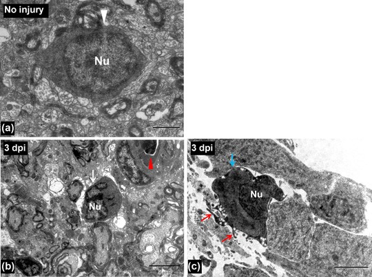
Figure Caption
Figure 3
(A) TEM image of an uninjured spinal cord showing a microglia (white arrowhead). (B) TEM image of a 3‐day post injured spinal cord showing a microglia (white arrow) near the injury site. Red arrowhead indicates a blood vessel. (C) TEM image of a 3‐day post injured spinal cord showing an activated microglia near the central canal (having finger‐like cytoplasmic projections, red arrows) of injured spinal cord (blue arrow). Nu, cell nucleus. Scale bar 1 μm (A), 5 μm (B), 2 μm (C)
Acknowledgments
This image is the copyrighted work of the attributed author or publisher, and
ZFIN has permission only to display this image to its users.
Additional permissions should be obtained from the applicable author or publisher of the image.
Full text @ Regeneration (Oxf)