
- Title
-
Teratogenic Effects of Topiramate in a Zebrafish Model
- Authors
- Lai, Y.H., Ding, Y.J., Moses, D., Chen, Y.H.
- Source
- Full text @ Int. J. Mol. Sci.
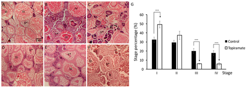
Under-maturation of oogenesis was shown in topiramate-treated female fish. Developmental status of oocyte in maternal fish was determined by Hematoxylin and eosin (H & E) staining. (A) Generally, the ovary was filled with mature oocytes in control fish in a high percentage; (B–F) however, a decreased percentage of mature oocytes was detected in different topiramate-treated female fish. Identification of the maturation stage was based on morphologies (I, primary growth stage; II, early cortical alveolus stage; II *, mid-cortical alveolus stage III, vitellogesis stage; IV, mature oocyte); (G) statistical analysis of oogenesis stages. Columns represent the number of cells at the four stages as percentages of the total counted (averages ± SD; *** p < 0.005; control n = 8; topiramate-treated n = 7). PHENOTYPE:
|
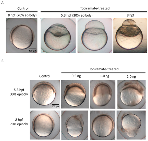
Embryonic epiboly deficiency in topiramate-treated offspring. (A) Embryonic development observation from control and topiramate-treated offspring. Morphology at 8 hpf in the control group showed normal epiboly progression. Embryos from topiramate-treated female fish were abnormally expressed during epiboly progression from 5.3 hpf; (B) embryonic development observation from control and topiramate-injected embryos at 5.3 and 8 hpf. Scale bar: 200 μm. |
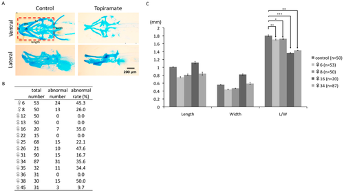
Cartilage development of offspring was impaired through maternal transmission of topiramate. (A) Ventral and lateral views of cartilage structure were detected by Alcian blue staining at 4 dpf larvae. Compared to the control group, there was a lack or shortage of cranial and pharyngeal development in topiramate-treated offspring; (B) embryonic number and abnormal rate were calculated from 14 female zebrafish; (C) length and width from individual maternal fish were measured and statistically analyzed. (averages ± SD; * p < 0.5; ** p < 0.01; *** p < 0.005; cb, ceratobranchial; ch, ceratohyal; ep, ethmoid plate; hs, hyosymplectic; M, Meckel’s cartilage; pq, palatoquadrate; Scale bar: 200 μm). |
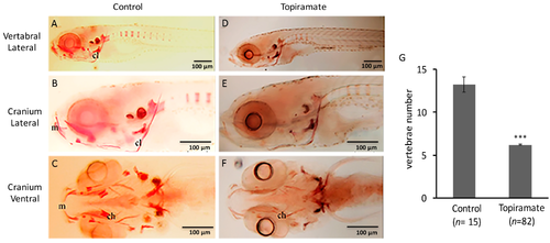
Bone development of offspring was impaired through maternal transmission of topiramate. (A–F) Ossification status of cranium and vertebral regions was detected by Alizarin red staining of zebrafish at 10 dpf; (G) Vertebrae numbers from offspring of control or topiramate-treated female zebrafish (♀25) (averages ± SD; *** p < 0.005; control n = 15; topiramate-treated n = 82). (ch, ceratohyal; cl, cleithrum; m, Meckel’s cartilage; Scale bar: 100 μm). |
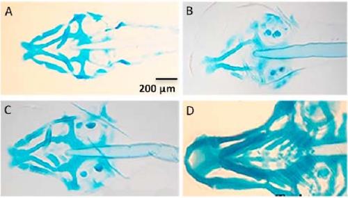
Craniofacial cartilage abnormalities upon diverse topiramte-treated offspring. |