
|
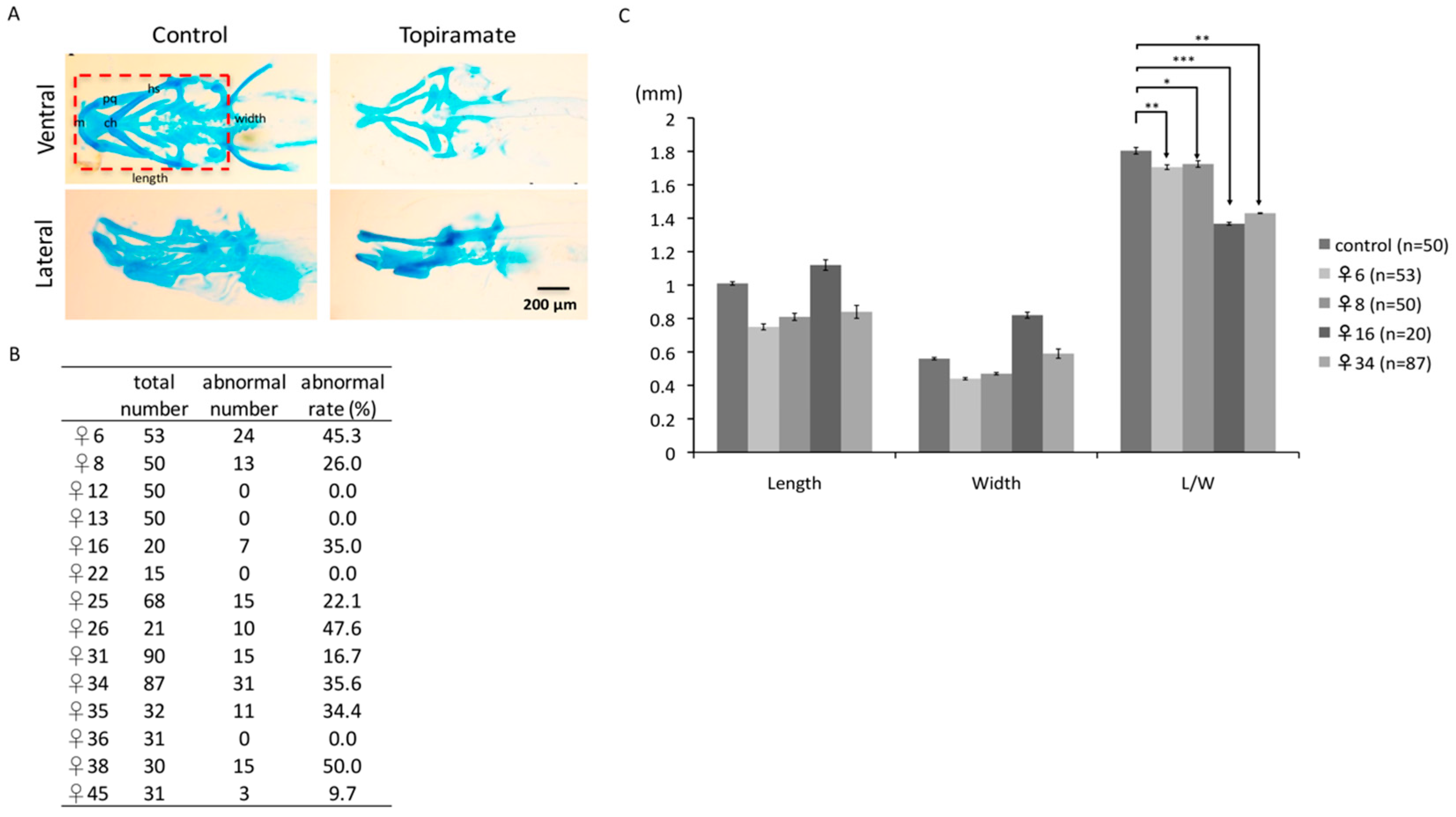
Fig. 3
Cartilage development of offspring was impaired through maternal transmission of topiramate. (A) Ventral and lateral views of cartilage structure were detected by Alcian blue staining at 4 dpf larvae. Compared to the control group, there was a lack or shortage of cranial and pharyngeal development in topiramate-treated offspring; (B) embryonic number and abnormal rate were calculated from 14 female zebrafish; (C) length and width from individual maternal fish were measured and statistically analyzed. (averages ± SD; * p < 0.5; ** p < 0.01; *** p < 0.005; cb, ceratobranchial; ch, ceratohyal; ep, ethmoid plate; hs, hyosymplectic; M, Meckel’s cartilage; pq, palatoquadrate; Scale bar: 200 μm).