
|
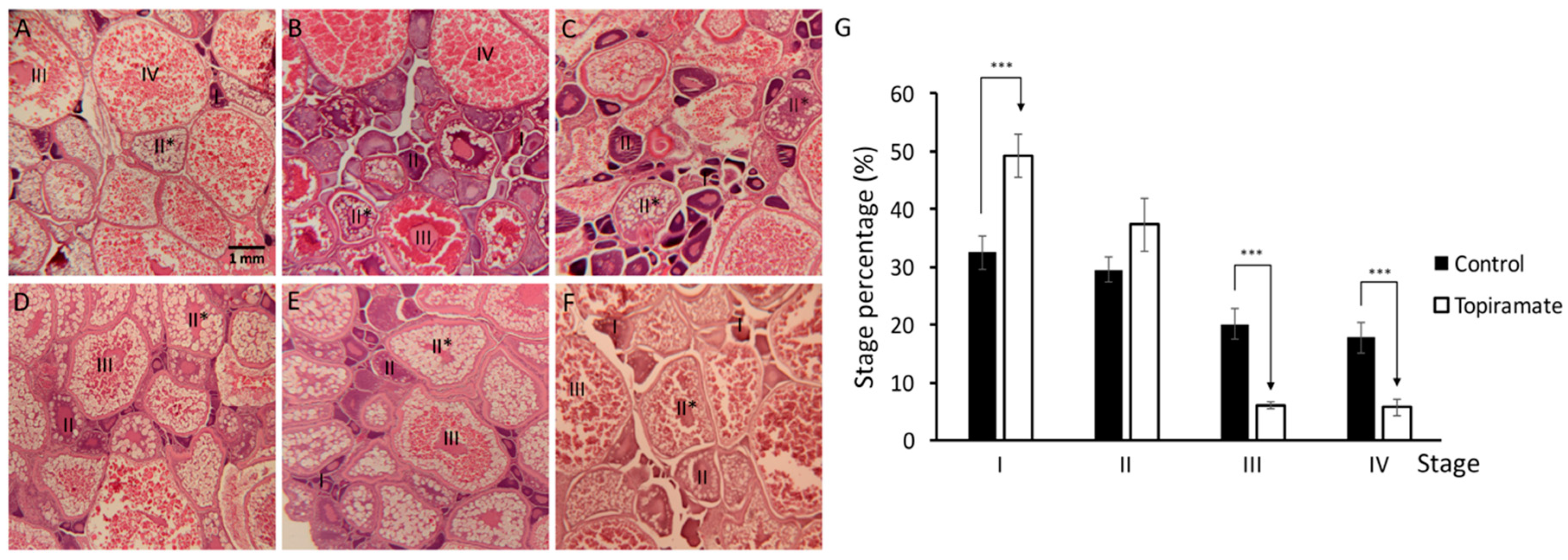
Fig. 1
Under-maturation of oogenesis was shown in topiramate-treated female fish. Developmental status of oocyte in maternal fish was determined by Hematoxylin and eosin (H & E) staining. (A) Generally, the ovary was filled with mature oocytes in control fish in a high percentage; (B–F) however, a decreased percentage of mature oocytes was detected in different topiramate-treated female fish. Identification of the maturation stage was based on morphologies (I, primary growth stage; II, early cortical alveolus stage; II *, mid-cortical alveolus stage III, vitellogesis stage; IV, mature oocyte); (G) statistical analysis of oogenesis stages. Columns represent the number of cells at the four stages as percentages of the total counted (averages ± SD; *** p < 0.005; control n = 8; topiramate-treated n = 7).