
- Title
-
Transmission Electron Microscopy of Zebrafish Spinal Motor Nerve Roots
- Authors
- Morris, A.D., Erisir, A., Criswell, S.J., Kucenas, S.
- Source
- Full text @ Dev. Dyn.
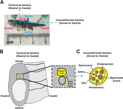
Spinal motor nerves in adult zebrafish. A: Image of an adult zebrafish: rostral to left, caudal to right, dorsal to top, and ventral to bottom. A dashed box identifies the trunk region of interest (ROI). Magenta dashed line and arrow identify the transverse sectioning orientation in a rostral-to-caudal direction, with 1 representing that the first section is most rostral and proceeds caudally. Blue dashed line and arrow identify the coronal/frontal sectioning orientation in a dorsal-to-ventral direction, with 1 representing that the first section is most dorsal and proceeds ventrally. B: Cartoon schematic of a transverse section of the trunk region, identifying the spinal cord and spinal motor nerves in yellow. The dashed box represents an inset with a higher-magnification view of the spinal cord and motor nerve roots. Blue arrow denotes ROI for coronal/frontal sectioning of a spinal motor nerve root. C: Cartoon schematic of a coronal/frontal cross-section of a spinal motor nerve. Myelinated axons (magenta) are surrounded by the endoneurium (orange). Several myelinated axons are ensheathed by the perineurium (green) to form a nerve fascicle, and several nerve fascicles are brought together into a nerve bundle by the epineurium (yellow). An artery (red) and vein (blue) are also denoted. |
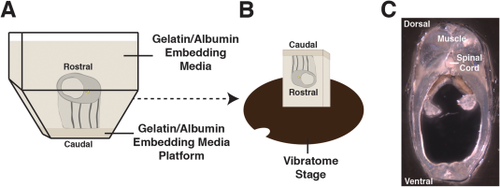
Vibrating blade microtome transverse sections of adult zebrafish trunk samples. A: Cartoon schematic portraying the dissected trunk region embedded in gelatin/albumin. B: Following embedding media solidification, the sample is removed from the mold and trimmed down using a razor blade. The sample is then mounted on the vibratome platform for sectioning. C: 200 µm transverse section of an adult zebrafish trunk region with dorsal to the top. |
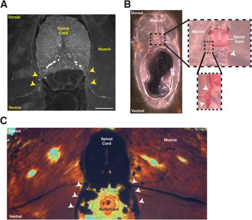
Locating spinal motor nerves in adult zebrafish transverse sections. A: Immunohistochemistry on floating sections using an antibody to tubulin. Yellow arrowheads identify the tubulin+ peripheral motor nerves. Scale bar = 100 µm. B: Black Gold II myelin stain to label the myelin sheath. The dotted box denotes the area enlarged in the right and bottom right images. White arrowheads identify the myelin sheath associated with peripheral motor axons. C: Osmium labeling identifies the myelin sheath associated with peripheral motor axons (white arrowheads). |
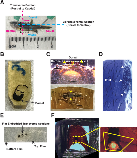
Embedding transverse sections for ultrathin sectioning in coronal/frontal and transverse orientations. A: Image of an adult zebrafish: rostral to left, caudal to right, dorsal to top, and ventral to bottom. A dashed box identifies the trunk ROI. Magenta dashed line and arrow identify transverse sectioning orientation in a rostral-to-caudal direction, with 1 representing the first section is most rostral and proceeds caudally. Blue dashed line and arrow identify coronal/frontal sectioning orientation in a dorsal-to-ventral direction, with 1 representing the first section is most dorsal and proceeds ventrally. B: Transverse section embedded within a flat mold, dorsal side oriented toward tip of mold to allow for coronal/frontal sections of the motor nerve. C: For coronal/frontal sections, a razor blade is used to remove the dorsal region of the sample. A trapezoid is created containing the ROI for ultrathin sectioning (Top: lateral view; bottom: top view). Yellow circle denotes notochord. Yellow arrowheads identify trapezoid. White dashed line and arrow identify coronal/frontal sectioning orientation in a dorsal-to-ventral direction, with 1 representing the first section is most dorsal and proceeds ventrally D: To confirm ROI is capturing a coronal/frontal cross-section of the motor nerves, 0.5 µm thick sections of a large area (several mm) are collected on a glass slide and stained with toluidine blue. Myelinated axons (a) are seen within the peripheral nerve (white arrowheads). Muscle (mu) is adjacent to the peripheral nerve. E: For transverse sections, samples are flat-embedded in EPON between two ACLAR films. F: Flat-embedded samples are excised from the film and re-embedded in a capsule. Polymerized EPON is removed to expose the sample (left), and a trapezoid ROI is identified (right). A trapezoid is created based on the identified ROI, and ultrathin sectioning in a transverse orientation is performed. |
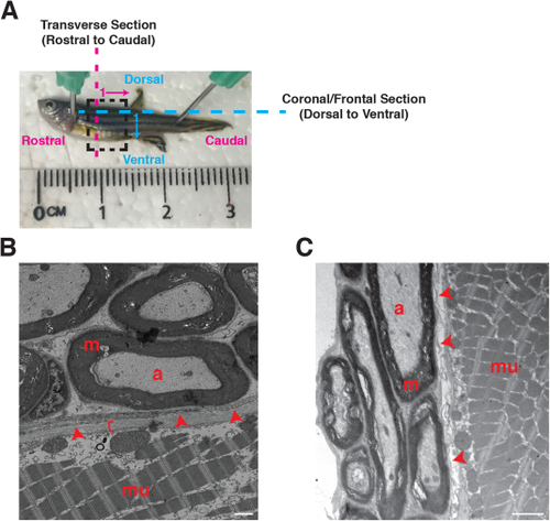
Ultrastructural evaluation of adult zebrafish motor nerves. A: Image of an adult zebrafish: rostral to left, caudal to right, dorsal to top, and ventral to bottom. A dashed box identifies the trunk ROI. Magenta dashed line and arrow identify transverse sectioning orientation in a rostral-to-caudal direction, with 1 representing the first section is most rostral and proceeds caudal. Blue dashed line and arrow identify coronal/frontal sectioning orientation in a dorsal-to-ventral direction, with 1 representing the first section is most dorsal and proceeds ventrally. B: Coronal/frontal view of an adult zebrafish motor nerve. C: Transverse view of an adult zebrafish motor nerve. a, axon; m, myelin; mu, muscle; arrowheads, perineurium; bracket, epineurium. Scale bar = 1 µm. |