
Fig. 4
- ID
- ZDB-FIG-180130-25
- Publication
- Morris et al., 2017 - Transmission Electron Microscopy of Zebrafish Spinal Motor Nerve Roots
- Other Figures
- All Figure Page
- Back to All Figure Page
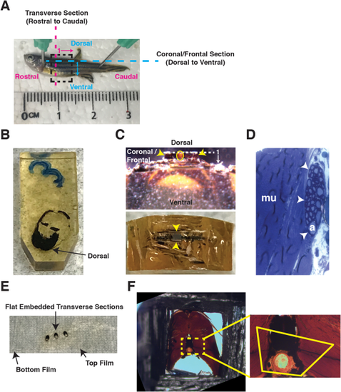
Embedding transverse sections for ultrathin sectioning in coronal/frontal and transverse orientations. A: Image of an adult zebrafish: rostral to left, caudal to right, dorsal to top, and ventral to bottom. A dashed box identifies the trunk ROI. Magenta dashed line and arrow identify transverse sectioning orientation in a rostral-to-caudal direction, with 1 representing the first section is most rostral and proceeds caudally. Blue dashed line and arrow identify coronal/frontal sectioning orientation in a dorsal-to-ventral direction, with 1 representing the first section is most dorsal and proceeds ventrally. B: Transverse section embedded within a flat mold, dorsal side oriented toward tip of mold to allow for coronal/frontal sections of the motor nerve. C: For coronal/frontal sections, a razor blade is used to remove the dorsal region of the sample. A trapezoid is created containing the ROI for ultrathin sectioning (Top: lateral view; bottom: top view). Yellow circle denotes notochord. Yellow arrowheads identify trapezoid. White dashed line and arrow identify coronal/frontal sectioning orientation in a dorsal-to-ventral direction, with 1 representing the first section is most dorsal and proceeds ventrally D: To confirm ROI is capturing a coronal/frontal cross-section of the motor nerves, 0.5 µm thick sections of a large area (several mm) are collected on a glass slide and stained with toluidine blue. Myelinated axons (a) are seen within the peripheral nerve (white arrowheads). Muscle (mu) is adjacent to the peripheral nerve. E: For transverse sections, samples are flat-embedded in EPON between two ACLAR films. F: Flat-embedded samples are excised from the film and re-embedded in a capsule. Polymerized EPON is removed to expose the sample (left), and a trapezoid ROI is identified (right). A trapezoid is created based on the identified ROI, and ultrathin sectioning in a transverse orientation is performed. |