
|
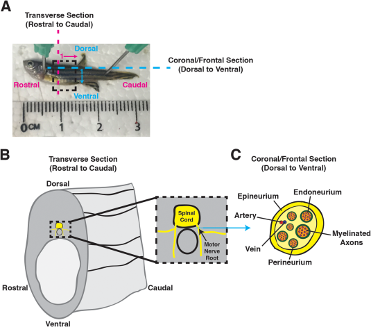
Fig. 1 Spinal motor nerves in adult zebrafish. A: Image of an adult zebrafish: rostral to left, caudal to right, dorsal to top, and ventral to bottom. A dashed box identifies the trunk region of interest (ROI). Magenta dashed line and arrow identify the transverse sectioning orientation in a rostral-to-caudal direction, with 1 representing that the first section is most rostral and proceeds caudally. Blue dashed line and arrow identify the coronal/frontal sectioning orientation in a dorsal-to-ventral direction, with 1 representing that the first section is most dorsal and proceeds ventrally. B: Cartoon schematic of a transverse section of the trunk region, identifying the spinal cord and spinal motor nerves in yellow. The dashed box represents an inset with a higher-magnification view of the spinal cord and motor nerve roots. Blue arrow denotes ROI for coronal/frontal sectioning of a spinal motor nerve root. C: Cartoon schematic of a coronal/frontal cross-section of a spinal motor nerve. Myelinated axons (magenta) are surrounded by the endoneurium (orange). Several myelinated axons are ensheathed by the perineurium (green) to form a nerve fascicle, and several nerve fascicles are brought together into a nerve bundle by the epineurium (yellow). An artery (red) and vein (blue) are also denoted.