
|
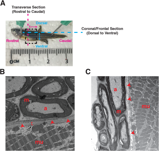
Fig. 5 Ultrastructural evaluation of adult zebrafish motor nerves. A: Image of an adult zebrafish: rostral to left, caudal to right, dorsal to top, and ventral to bottom. A dashed box identifies the trunk ROI. Magenta dashed line and arrow identify transverse sectioning orientation in a rostral-to-caudal direction, with 1 representing the first section is most rostral and proceeds caudal. Blue dashed line and arrow identify coronal/frontal sectioning orientation in a dorsal-to-ventral direction, with 1 representing the first section is most dorsal and proceeds ventrally. B: Coronal/frontal view of an adult zebrafish motor nerve. C: Transverse view of an adult zebrafish motor nerve. a, axon; m, myelin; mu, muscle; arrowheads, perineurium; bracket, epineurium. Scale bar = 1 µm.