
- Title
-
Regulatory evolution of Tbx5 and the origin of paired appendages
- Authors
- Adachi, N., Robinson, M., Goolsbee, A., Shubin, N.H.
- Source
- Full text @ Proc. Natl. Acad. Sci. USA
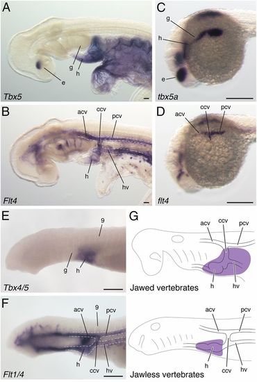
Comparison of Tbx5 cognate genes in vertebrates. Skate Tbx5 (A) and Flt4 (B) expression at stage 23. Zebrafish tbx5a (C) and flt4 (D) expression at 22 hpf. Sea lamprey Tbx4/5 (E) and Flt1/4 (F) expression at stage 24. (G) A depiction of the Tbx5 expression domain (purple) relative to embryonic components in jawed and jawless vertebrates. acv, anterior cardinal vein; ccv, common cardinal vein; e, eye; g, gills; h, heart; hv, hepatocardiac vein; pcv, posterior cardinal vein; 9, somite 9. (Scale bars: 200 µm.) |
Gar CNS12 drives GFP expression in the pectoral fin of zebrafish. Brightfield (A, D, and G) and fluorescent (B, E, and H) images of gar CNS12 transgenic zebrafish. Lateral views are in A-C and dorsal views are in D-I. GFP expression is found in the dorsal part of the eye and pectoral fin field at 24 hpf (A, B, D, and E), and in the common cardinal vein (arrowhead) and pectoral fin bud at 36 hpf (G and H). Dotted lines outline the pectoral fin. Zebrafish tbx5a in situ hybridization at 24 hpf (C and F) and at 36 hpf (I). (Scale bars: 200 µm.) |
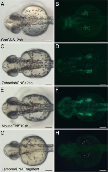
Brightfield (A, C, and E) and fluorescent (B, D, and F) images of CNS12sh transgenic zebrafish from dorsal view at 36 hpf. Gar (A and B), zebrafish (C and D), and mouse (E and F) CNS12sh drive GFP expression in the pectoral fin bud. (G and H) Transgenic zebrafish with Japanese lamprey DNA fragment aligned to jawed vertebrate CNS12 at 36 hpf. Dotted lines outline the pectoral fin. (Scale bars: 200 µm.) EXPRESSION / LABELING:
|
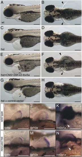
tbx5a driven by CNS12sh rescues pectoral fin formation in hst. Wild-type (A and B) and hst (CH) at 3 dpf. Lateral views (A, C, E, and G) and dorsal views (B, D, F, and H). Black arrowheads indicate pectoral fins. fgf8a (I and L), fgf10a (J and M), and col2a1a (K and N) in situ hybridization in the wild-type (I-K) and rescued hst (LN) at 3 dpf. Dotted lines outline the pectoral fin and white arrowheads indicate the scapulocoracoid. (Scale bars: A-H, 200 µm; I-N, 100 µm.) |
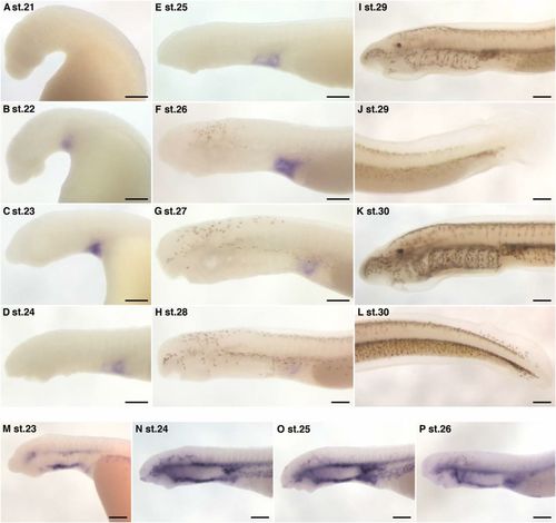
Tbx4/5 and Flt1/4 expression in sea lamprey embryos. Tbx4/5 (AL) and Flt1/4 (MP) expression in developing sea lampreys. (A) Tbx4/5 was undetectable at stage 21. (B) Tbx4/5 is initially detected in the heart primordium at stage 22. (CF) Tbx4/5 is expressed in the heart. The posterior border of Tbx4/5 expression and heart domain are not clear due to the premature development of heart and cardinal veins at these stages. (G and H) The heart expression of Tbx4/5 becomes barely detectable at stages 27 and 28. (I-L) Tbx4/5 is undetectable in the heart at stages 29 and 30. The expression is never found in the median fin (J and L). Tbx4/5 expression was not found in the dorsal portion of the eye in sea lamprey embryos. (M) Flt1/4 was detectable in the dorsal and ventral aortas, but not in the cardinal veins at stage 23. (N and O) Flt1/4 was detected in the cardinal veins at stages 24 and 25. (P) Flt1/4 was weakly expressed in the cardinal veins at stage 26. (Scale bars: 200 µm.) |
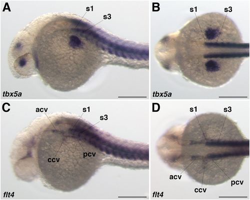
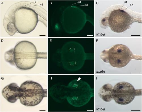
tbx5a and flt4 expression in zebrafish. tbx5a (A and B) and flt4 (C and D) expression at 24 hpf of zebrafish embryos. Dorsolateral views (A and C) and dorsal views (B and D). tbx5a is expressed next to somites 1-4. The posterior half of the common cardinal vein labeled by flt4 is located next to somite 1. acv, anterior cardinal vein; ccv, common cardinal vein; pcv, posterior cardinal vein; s1, somite 1; s3, somite 3. (Scale bars: 200 µm.) |
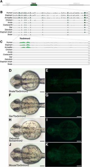
Sequence alignment of Tbx5 coding region and Tbx5 intron2 transgenic zebrafish. (A) A schematic representation of human TBX5 locus. (B) mVISTA analysis around Tbx5 using the mouse sequence as base. (C) Enlargement of Tbx5 intron2. Conserved sequences are indicated as peaks. Brightfield (D, F, H, and J) and fluorescent (E, G, I, and K) images of Tbx5 intron2 transgenic zebrafish. All images are from dorsal view at 36 hpf. Skate (D and E), gar (F and G), zebrafish (H and I) and mouse (J and K) Tbx5 intron2 never drive GFP signal in the pectoral fin bud. (Scale bars: 200 µm.) |
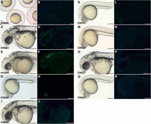
Transgenic zebrafish with DNA fragment around Tbx5 locus. Brightfield (A, C, E, G, I, K, M, O, and Q) and fluorescent (B, D, F, H, J, L, N, P, and R) images. (A and B) CNS59 transgenic zebrafish at 9 hpf. One stable line is screened. (C and D) CNS63 transgenic zebrafish at 36 hpf: one line. (E and F) Tbx5 intron5 transgenic zebrafish at 36 hpf: seven lines. (G and H) CNS58 transgenic zebrafish at 24 hpf: two lines. (I and J) CNS23 transgenic zebrafish at 36 hpf: six lines. (K and L) CNS11 transgenic zebrafish at 24 hpf: one line. (M and N) CNS26 transgenic zebrafish at 36 hpf: one line. (O and P) CNS27 transgenic zebrafish at 36 hpf: one line. (Q and R) CNS65 transgenic zebrafish at 24 hpf: two lines. (Scale bars: 200 µm.) |
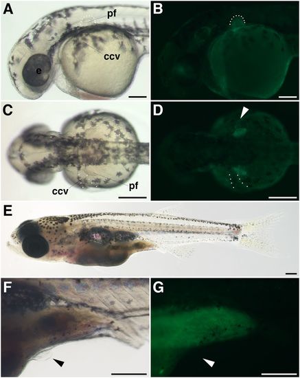
GFP expression pattern of gar CNS12 transgenic zebrafish. Brightfield (A, C, E, and F) and fluorescent (B, D, and G) images of gar CNS12 transgenic zebrafish. Lateral views in (A, B, and EG) and dorsal views in (C and D). (A and B) GFP expressions are found in the dorsal eye and pectoral fin bud at 36 hpf. Dotted lines outline the fin bud, in which GFP signals are detected in the mesenchyme. (C and D) GFP are expressed on the posterior edge of the ccv (dotted lines and arrowhead). (E) A 4-wk-old juvenile of the transgenic fish. (F) The juvenile has the pelvic fin bud (black arrowhead). (G) The fin bud does not show GFP signals (white arrowhead). A green emission is detected in the digestive tract. ccv, common cardinal vein; e, eye; pf, pectoral fin. (Scale bars: A-D, 200 µm; E-G, 500 µm.) |
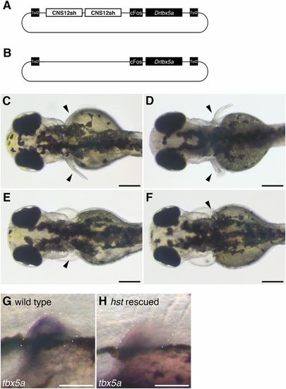
Various phenotypes of pectoral fins in rescued hst. Schematic of vectors used in the rescue (A) and control (B) experiments. Dorsal views of rescued hst zebrafish at 3 dpf. Arrowheads indicate pectoral fins. (C) The right fin, almost identical to the wild-type fin, is well developed compared with the left one. (D) Likewise in C, the right fin is bigger than the left one, but both right and left fins are less developed compared with the wild-type fin. (E) A small left fin is rescued. (F) A small right fin is rescued. (G and H) tbx5a in situ hybridization in the wild-type (G) and rescued hst (H) at 3 dpf. Dotted lines outline the fin bud. cFos, cFos minimal promoter; CNS12sh, CNS12 short; Drtbx5a, Danio rerio tbx5a coding sequence; Tol2, Tol2 transposon sequence. (Scale bars: C-F, 200 µm; G and H, 100 µm.) |