
- Title
-
Functional bone histology of zebrafish reveals two types of endochondral ossification, different types of osteoblast clusters and a new bone type
- Authors
- Weigele, J., Franz-Odendaal, T.A.
- Source
- Full text @ J. Anat.
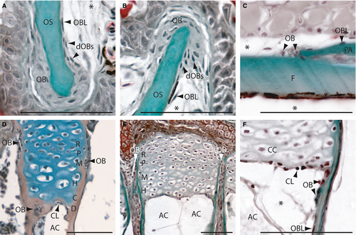
Intramembranous (A-C) and endochondral (D-F) ossification in zebrafish on longitudinal (A,B,E,F) and transversal (C,D) Mallory′s trichrome-stained sections. (A,B) Osteoblasts cluster type I (OB) at the anterior premaxillary (A) and dentary (B). (C) Osteoblast clusters type II at the coronal suture of the frontal and parietal bone. (D) Section of an epiphysial plate of a radial located in the pectoral girdle, showing the epiphysial growth plate of type I endochondral bones. (E,F) Sections of the epiphysial plate with type II ossification located in the hypurals of the caudal fin. (F) Detail of the active chondroclast zone. AC, adipocyte; C, zone of calcification; CC, chondrocyte; CL, chondroclasts; D, zone of cartilage degradation; dOBs, differentiating osteoblasts; F, frontal bone; H, zone of hypertrophic chondrocytes; M, zone of maturation; OB; osteoblast; OBL, osteoblast-like cell; OS, osseus (mineralized bone); P, zone of proliferation; PA, parietal bone; R, zone of reserve cartilage. *Artefact. Scale bars: 50 µm. |
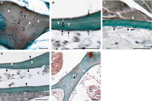
Histology of adult zebrafish compact bones on transverse Mallory′s Trichrome-stained sections. (A) Section of the cellular opercular bone. Black arrowheads are indicating round osteocytes with oval, round or atypical formed nuclei and white arrowheads elongated osteocytes with spindle-shaped nuclei. (B) Transition from osteoblast-like cells (1) through intermediary stages to partial bone entrapment of cells (2-4), and finally to full entrapment as osteocytes (5; black arrowheads). White arrowheads mark the cellular processes of an osteocyte. (C) Section of an acellular bone of the opercle. Arrowheads indicate osteoblast-like cells at the surfaces. (D) Section of two branchiostegal rays with cellular (upper) and acellular (lower) parts. Black arrowheads indicate osteoblast-like cells and white arrowheads, osteocytes. (E) Section of the lateral ethmoid with an osteon. Black arrowhead is indicating the Haversian canal and the white arrowhead marks the cement line to the surrounding bone tissue. Scale bars: 25 µm. |
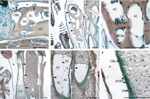
Histology of adult zebrafish spongy bones (A-C) and tubular bones (D-F) on transversal (A-D, F) and longitudinal Mallory′s Trichrome-stained sections (E). (A) Section of the pectoral girdle and the ceratohyal bone (B, arrowheads). (C) Detail of a spongy bone (the ceratohyal), with osteoblasts, osteoclasts, osteoblast-like cells and adipose cells. (D) Section of the tubular hyomandibular (arrowheads). (E) Section of two hypuralia, arrowheads are indicating the osseus. (F) Detail of the hyomandibular, with osteoblasts (bone collar cells) at the outer surface developing to osteoblast-like cells. White arrowheads indicate the developing osseus. AC, adipocyte; CA, Cartilage; dOBs, differentiating osteoblasts; OB, osteoblast; OBL, osteoblast-like cell; OC, osteoclast; OS, osseus. *Artefact. Scale bars: 50 µm. |
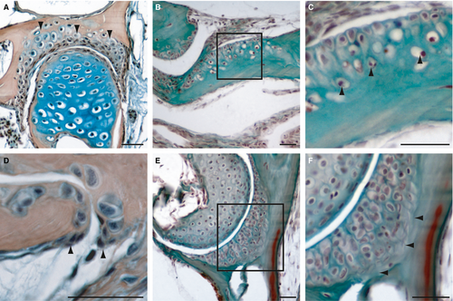
Histology of adult zebrafish chondroid bones on transversal (A, D-F) and longitudinal sections (B,C). (A) Mallory′s Trichrome and (B-F) HBQ staining. (A) Section of the hyomandibular joint, arrowheads are indicating the chondroid bone. (B,C) Sections of the premaxillary. (C) Detail of the transition to normal bone, arrowheads are indicating bone tissue entrapped chondrocytes. (D) Detail of the hyomandibular-opercle joint edge, arrowheads are indicating osteoblast clusters of type II. (E,F) Section of the hyomandibular-opercle joint, detail (F) shows a collagen-rich zone (arrowheads) between the chondroid cartilage and the bone tissue. Scale bars: 25 µm. |