
Fig. 4
- ID
- ZDB-FIG-160714-7
- Publication
- Weigele et al., 2016 - Functional bone histology of zebrafish reveals two types of endochondral ossification, different types of osteoblast clusters and a new bone type
- Other Figures
- All Figure Page
- Back to All Figure Page
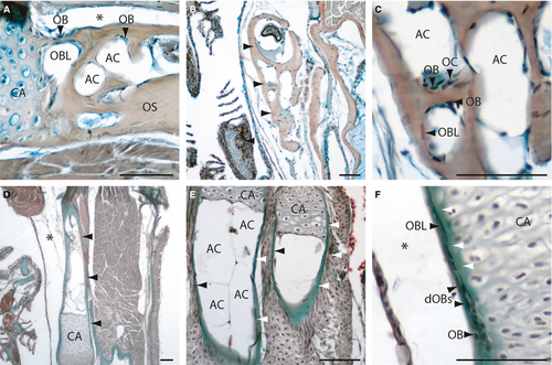
Histology of adult zebrafish spongy bones (A-C) and tubular bones (D-F) on transversal (A-D, F) and longitudinal Mallory′s Trichrome-stained sections (E). (A) Section of the pectoral girdle and the ceratohyal bone (B, arrowheads). (C) Detail of a spongy bone (the ceratohyal), with osteoblasts, osteoclasts, osteoblast-like cells and adipose cells. (D) Section of the tubular hyomandibular (arrowheads). (E) Section of two hypuralia, arrowheads are indicating the osseus. (F) Detail of the hyomandibular, with osteoblasts (bone collar cells) at the outer surface developing to osteoblast-like cells. White arrowheads indicate the developing osseus. AC, adipocyte; CA, Cartilage; dOBs, differentiating osteoblasts; OB, osteoblast; OBL, osteoblast-like cell; OC, osteoclast; OS, osseus. *Artefact. Scale bars: 50 µm. |