
Fig. 3
- ID
- ZDB-FIG-160714-6
- Publication
- Weigele et al., 2016 - Functional bone histology of zebrafish reveals two types of endochondral ossification, different types of osteoblast clusters and a new bone type
- Other Figures
- All Figure Page
- Back to All Figure Page
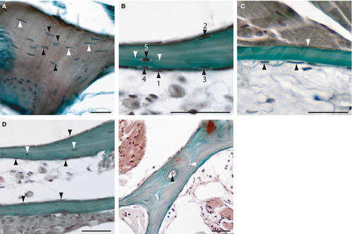
Histology of adult zebrafish compact bones on transverse Mallory′s Trichrome-stained sections. (A) Section of the cellular opercular bone. Black arrowheads are indicating round osteocytes with oval, round or atypical formed nuclei and white arrowheads elongated osteocytes with spindle-shaped nuclei. (B) Transition from osteoblast-like cells (1) through intermediary stages to partial bone entrapment of cells (2-4), and finally to full entrapment as osteocytes (5; black arrowheads). White arrowheads mark the cellular processes of an osteocyte. (C) Section of an acellular bone of the opercle. Arrowheads indicate osteoblast-like cells at the surfaces. (D) Section of two branchiostegal rays with cellular (upper) and acellular (lower) parts. Black arrowheads indicate osteoblast-like cells and white arrowheads, osteocytes. (E) Section of the lateral ethmoid with an osteon. Black arrowhead is indicating the Haversian canal and the white arrowhead marks the cement line to the surrounding bone tissue. Scale bars: 25 µm. |