
|
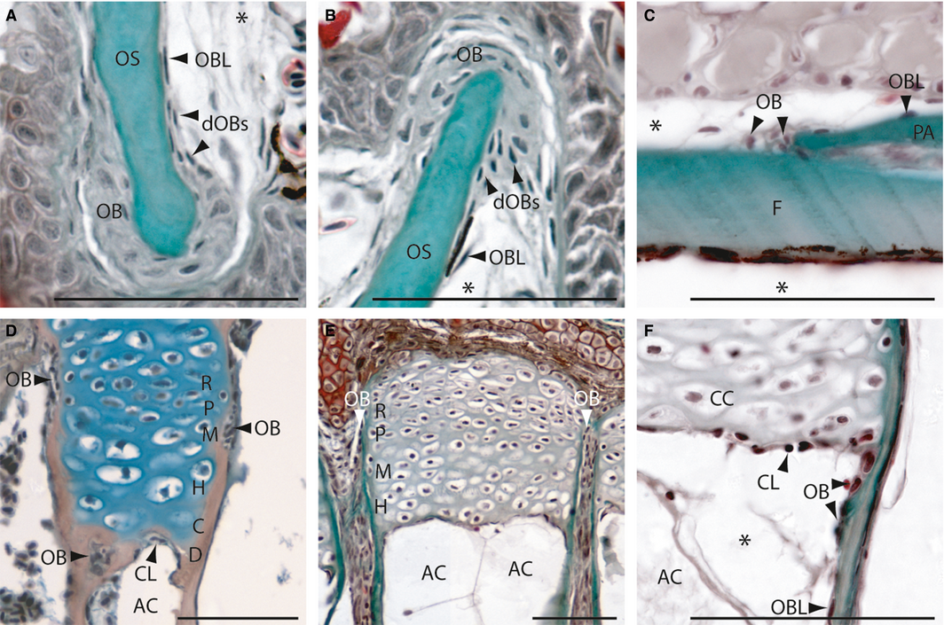
Fig. 1
Intramembranous (A-C) and endochondral (D-F) ossification in zebrafish on longitudinal (A,B,E,F) and transversal (C,D) Mallory′s trichrome-stained sections. (A,B) Osteoblasts cluster type I (OB) at the anterior premaxillary (A) and dentary (B). (C) Osteoblast clusters type II at the coronal suture of the frontal and parietal bone. (D) Section of an epiphysial plate of a radial located in the pectoral girdle, showing the epiphysial growth plate of type I endochondral bones. (E,F) Sections of the epiphysial plate with type II ossification located in the hypurals of the caudal fin. (F) Detail of the active chondroclast zone. AC, adipocyte; C, zone of calcification; CC, chondrocyte; CL, chondroclasts; D, zone of cartilage degradation; dOBs, differentiating osteoblasts; F, frontal bone; H, zone of hypertrophic chondrocytes; M, zone of maturation; OB; osteoblast; OBL, osteoblast-like cell; OS, osseus (mineralized bone); P, zone of proliferation; PA, parietal bone; R, zone of reserve cartilage. *Artefact. Scale bars: 50 µm.