
|
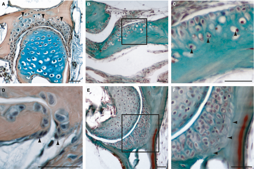
Fig. 5
Histology of adult zebrafish chondroid bones on transversal (A, D-F) and longitudinal sections (B,C). (A) Mallory′s Trichrome and (B-F) HBQ staining. (A) Section of the hyomandibular joint, arrowheads are indicating the chondroid bone. (B,C) Sections of the premaxillary. (C) Detail of the transition to normal bone, arrowheads are indicating bone tissue entrapped chondrocytes. (D) Detail of the hyomandibular-opercle joint edge, arrowheads are indicating osteoblast clusters of type II. (E,F) Section of the hyomandibular-opercle joint, detail (F) shows a collagen-rich zone (arrowheads) between the chondroid cartilage and the bone tissue. Scale bars: 25 µm.