
- Title
-
Galanin regulates blood glucose level in the zebrafish: a morphological and functional study
- Authors
- Podlasz, P., Jakimiuk, A., Chmielewska-Krzesinska, M., Kasica, N., Nowik, N., Kaleczyc, J.
- Source
- Full text @ Histochem. Cell Biol.

Whole-mount fluorescence in situ hybridization of galanin expression of 3 day post-fertilization (dpf) zebrafish larvae. Arrows point to galaninergic neurons in the autonomic ganglion. E eye, I intestine, Rho rhombencephalon, Y yolk. Scale bar 100 µm |
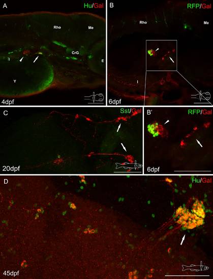
Whole-mount immunofluorescence staining of the zebrafish larvae (a, b) and juvenile zebrafish gut (c, d) using antibody against galanin (a-d) and neuronal marker Hu (a, d), somatostatin (Sst) (c). Red fluorescence protein (RFP) marked mnx1+ population of β cells (b, b′). Arrows show galaninergic neurons in the autonomic ganglion. In the primary islet of the endocrine pancreas, galanin-IR fibers formed a very dense network (arrowheads). CrG cranial ganglia, E eye, I intestine, Me mesencephalon, Rho rhombencephalon, Y yolk. Scale bars 100 µm |
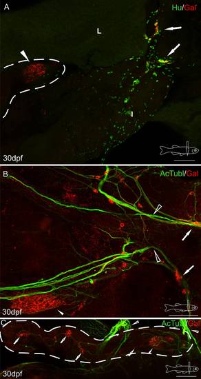
Whole-mount immunofluorescence staining of the 30 dpf zebrafish gut using antibody against galanin (a-c), neuronal marker Hu (a), and acetylated tubulin (b, c). Arrows point to galaninergic neurons in the autonomic ganglion, and the arrowhead shows a very dense network of galanin-IR fibers in the primary islet of the endocrine pancreas. The secondary islets (hollow arrows, c), the vagus nerve (hollow arrowheads, b, c) and its associated galanin-IR neurons are also visible. Dashed line indicates the pancreas (a, c). I intestine, L liver. Scale bars 100 µm |
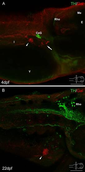
Whole-mount immunofluorescence staining of the 4 dpf zebrafish larvae (a), 22 dpf juvenile zebrafish (b) using antibody against galanin and adrenergic marker-tyrosine hydroxylase (TH). Arrow points to galaninergic neurons in autonomic ganglia. Arrowheads show the primary islet of the endocrine pancreas. Galanin-IR cells and fibers did not express immunoreactivity to TH. However, very intensely stained TH-IR neurons were found in the celiac ganglion (a) and in the brain (b). CeG celiac ganglion, E eye, I intestine, Me mesencephalon, Rho rhombencephalon. Scale bars 100 µm |
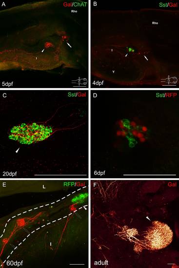
Whole-mount immunofluorescence staining of the zebrafish larvae (a, b, d), juvenile (c, e), and adult (f) guts using antibody against galanin (a-c, e-f), cholinergic marker-choline acetyltransferase (ChAT) (a), δ cell marker somatostatin (b-d). RFP marked mnx1+ population of β cells in the transgenic line of the zebrafish (d, e). Arrows point to galaninergic neurons in autonomic ganglia. Arrowheads show the primary islet of the endocrine pancreas, and hollow arrows point to the secondary islets. a There was no ChAT immunoreactivity in the cells and fibers associated with the pancreas and intestine. ChAT-IR neurons were observed only in the brain and spinal cord (not shown). Galanin-IR fibers were also visible, projecting from the autonomic ganglion to the intestine. c, d Pancreatic islets at a higher magnification. Somatostatin did not colocalize with β cell marker (d). Delta cells were very intensely supplied by galaninergic fibers (c, d). Note that the secondary islets did not contain RFP+ cells until 60 dpf (e). Dashed line indicates the pancreas. I intestine, L liver, Rho rhombencephalon. Scale bars 100 µm, except d = 50 µm |
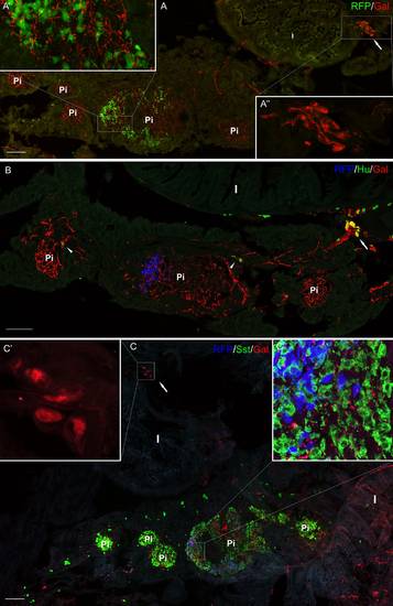
Section from the adult pancreas stained with antibody against galanin (a-c) and Hu (b), and somatostatin (c). RFP marked mnx1+ population of β cells in the transgenic line of the zebrafish (a-c). RFP+ cells were present mostly only in one (primary) islet, whereas somatostatin-positive cells were located in all the islets (a-c). Ganglion containing galanin-IR neurons is visible outside the pancreatic tissue, close to the intestinal wall (a, a′′, c, c′). Neurons inside the pancreatic tissue are also visible; however, they were mostly galanin-negative (arrowheads, b). Galanin-IR fibers richly supplied pancreatic islets (c). I intestine, Pi pancreatic islet. Scale bars 100 µm |