
Image
|
Figure Caption
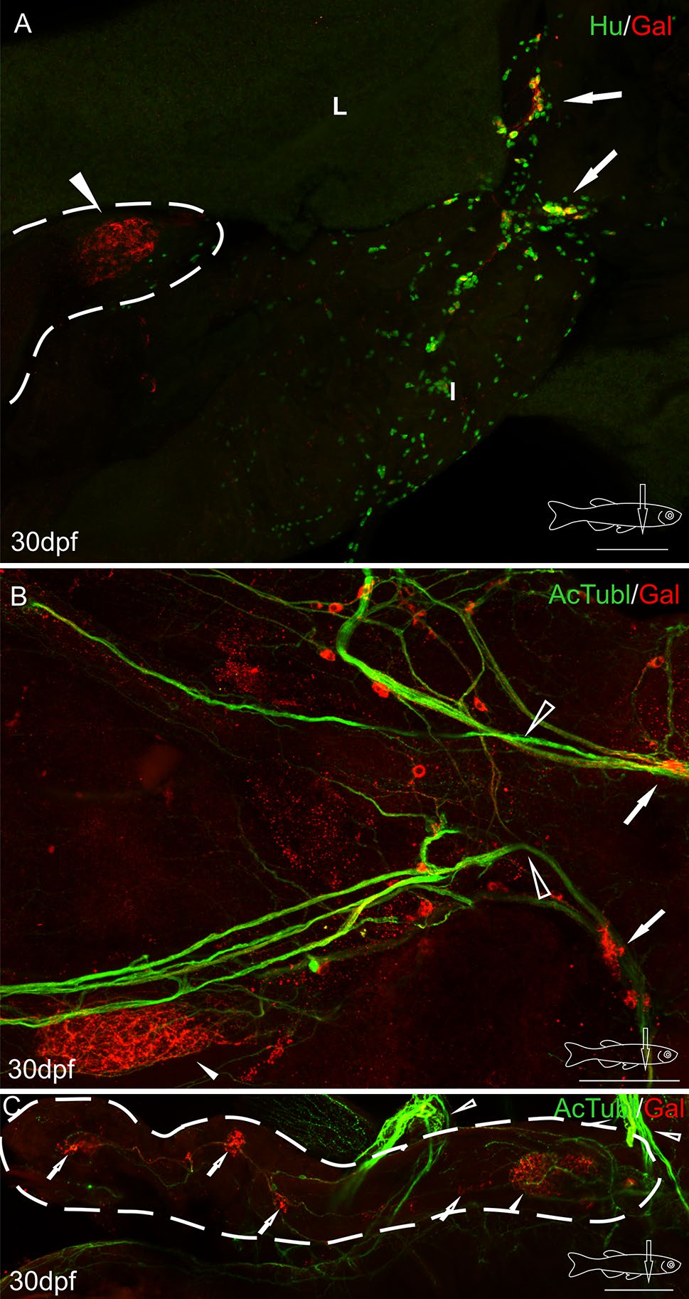
Fig. 3
Whole-mount immunofluorescence staining of the 30 dpf zebrafish gut using antibody against galanin (a-c), neuronal marker Hu (a), and acetylated tubulin (b, c). Arrows point to galaninergic neurons in the autonomic ganglion, and the arrowhead shows a very dense network of galanin-IR fibers in the primary islet of the endocrine pancreas. The secondary islets (hollow arrows, c), the vagus nerve (hollow arrowheads, b, c) and its associated galanin-IR neurons are also visible. Dashed line indicates the pancreas (a, c). I intestine, L liver. Scale bars 100 µm
Acknowledgments
This image is the copyrighted work of the attributed author or publisher, and
ZFIN has permission only to display this image to its users.
Additional permissions should be obtained from the applicable author or publisher of the image.
Full text @ Histochem. Cell Biol.