
Fig. 6
- ID
- ZDB-FIG-160526-15
- Publication
- Podlasz et al., 2016 - Galanin regulates blood glucose level in the zebrafish: a morphological and functional study
- Other Figures
- All Figure Page
- Back to All Figure Page
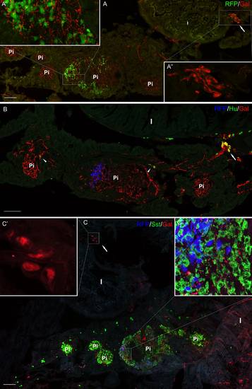
Section from the adult pancreas stained with antibody against galanin (a-c) and Hu (b), and somatostatin (c). RFP marked mnx1+ population of β cells in the transgenic line of the zebrafish (a-c). RFP+ cells were present mostly only in one (primary) islet, whereas somatostatin-positive cells were located in all the islets (a-c). Ganglion containing galanin-IR neurons is visible outside the pancreatic tissue, close to the intestinal wall (a, a′′, c, c′). Neurons inside the pancreatic tissue are also visible; however, they were mostly galanin-negative (arrowheads, b). Galanin-IR fibers richly supplied pancreatic islets (c). I intestine, Pi pancreatic islet. Scale bars 100 µm |