
FIGURE
Fig. 2
- ID
- ZDB-FIG-160526-11
- Publication
- Podlasz et al., 2016 - Galanin regulates blood glucose level in the zebrafish: a morphological and functional study
- Other Figures
- All Figure Page
- Back to All Figure Page
Fig. 2
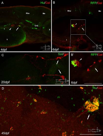
Whole-mount immunofluorescence staining of the zebrafish larvae (a, b) and juvenile zebrafish gut (c, d) using antibody against galanin (a-d) and neuronal marker Hu (a, d), somatostatin (Sst) (c). Red fluorescence protein (RFP) marked mnx1+ population of β cells (b, b′). Arrows show galaninergic neurons in the autonomic ganglion. In the primary islet of the endocrine pancreas, galanin-IR fibers formed a very dense network (arrowheads). CrG cranial ganglia, E eye, I intestine, Me mesencephalon, Rho rhombencephalon, Y yolk. Scale bars 100 µm |
Expression Data
Expression Detail
Antibody Labeling
Phenotype Data
Phenotype Detail
Acknowledgments
This image is the copyrighted work of the attributed author or publisher, and
ZFIN has permission only to display this image to its users.
Additional permissions should be obtained from the applicable author or publisher of the image.
Full text @ Histochem. Cell Biol.