
|
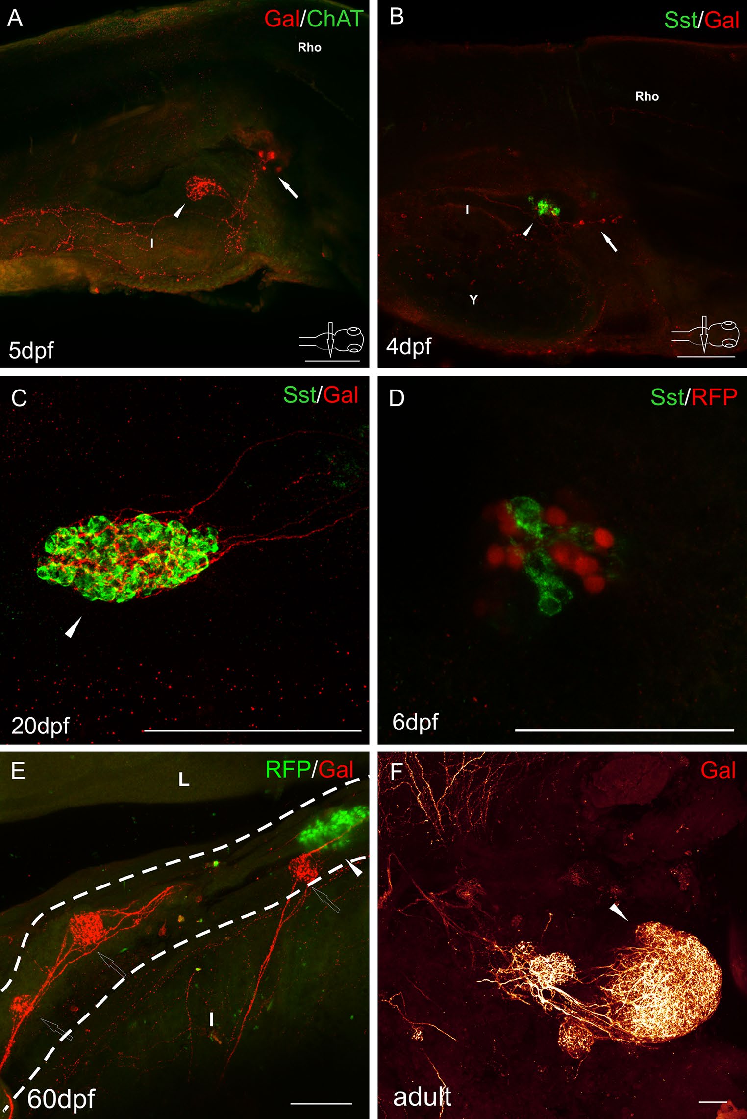
Fig. 5
Whole-mount immunofluorescence staining of the zebrafish larvae (a, b, d), juvenile (c, e), and adult (f) guts using antibody against galanin (a-c, e-f), cholinergic marker-choline acetyltransferase (ChAT) (a), δ cell marker somatostatin (b-d). RFP marked mnx1+ population of β cells in the transgenic line of the zebrafish (d, e). Arrows point to galaninergic neurons in autonomic ganglia. Arrowheads show the primary islet of the endocrine pancreas, and hollow arrows point to the secondary islets. a There was no ChAT immunoreactivity in the cells and fibers associated with the pancreas and intestine. ChAT-IR neurons were observed only in the brain and spinal cord (not shown). Galanin-IR fibers were also visible, projecting from the autonomic ganglion to the intestine. c, d Pancreatic islets at a higher magnification. Somatostatin did not colocalize with β cell marker (d). Delta cells were very intensely supplied by galaninergic fibers (c, d). Note that the secondary islets did not contain RFP+ cells until 60 dpf (e). Dashed line indicates the pancreas. I intestine, L liver, Rho rhombencephalon. Scale bars 100 µm, except d = 50 µm