
- Title
-
Transcription factors involved in retinogenesis are co-opted by the circadian clock following photoreceptor differentiation
- Authors
- Laranjeiro, R., Whitmore, D.
- Source
- Full text @ Development
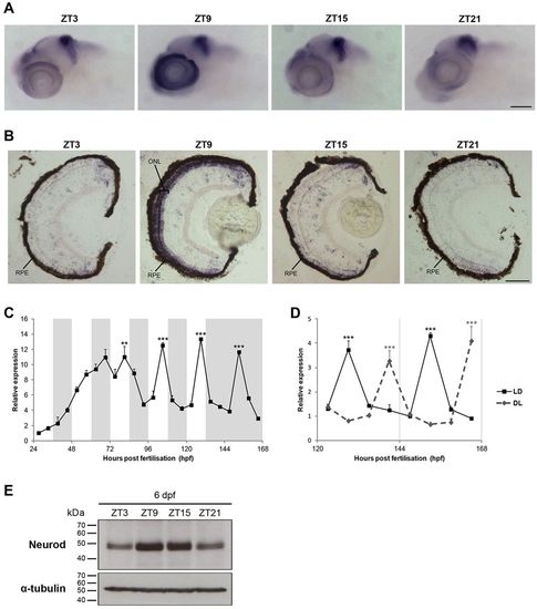
neurod rhythmic expression is restricted to retinal photoreceptors. (A,B) Representative images at four time points of neurod WISH on 6 dpf larvae (A) and of in situ hybridisation for neurod on 6 dpf larval eye sections (B). ZT, zeitgeber time; RPE, retinal pigment epithelium; ONL, outer nuclear layer. (C) qPCR analysis of neurod expression in zebrafish larvae raised on an LD cycle until 6dpf and then transferred to DD on day 7 (n=3). White and grey backgrounds represent light and dark phases, respectively. (D) qPCR analysis of neurod expression in 6-7dpf larvae raised on an LD cycle or a DL cycle (n=3). Statistically significant differences between the expression peak and trough on each day (Fisher′s LSD test) are indicated: **P<0.01, ***P<0.001. Error bars indicate s.e.m. (E) Representative western blots of Neurod and α-tubulin expression on 6dpf larvae at four time points. Scale bars: 100μm in A; 50μm in B. |
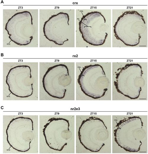
Circadian expression of retinal transcription factors in differentiated photoreceptors. (A-C) Representative images of in situ hybridisation for crx (A), rx2 (B) and nr2e3 (C) on 6dpf larval eye sections at four time points. RPE, retinal pigment epithelium; INL, inner nuclear layer; ONL, outer nuclear layer. Scale bars: 50μm. |
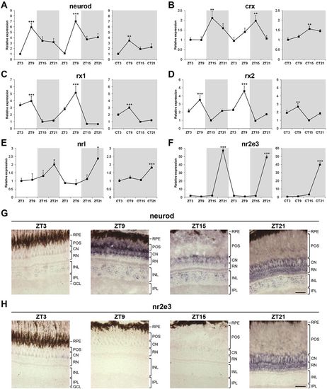
Circadian expression of retinal transcription factors is maintained in adult zebrafish. (A-F) qPCR analysis of neurod (A), crx (B), rx1 (C), rx2 (D), nrl (E) and nr2e3 (F) expression in adult eyes over two days of an LD cycle and one subsequent day of DD (n=3-4). Zeitgeber time (ZT) or circadian time (CT) indicates the four time points analysed per day. White and grey backgrounds represent light and dark phases, respectively. Statistically significant differences between the expression peak and trough on each day (Fisher′s LSD test) are indicated: *P<0.05, **P<0.01, ***P<0.001. Error bars indicate s.e.m. (G,H) Representative images of in situ hybridisation for neurod (G) and nr2e3 (H) on adult eye sections at four time points. Note that expression in the photoreceptors at ZT21 is restricted to rods for both neurod and nr2e3. The signal present in the layer of cone nuclei corresponds to rod inner segments that connect the rod nuclei to the rod outer segments. RPE, retinal pigment epithelium; POS, photoreceptor outer segments; CN, cone nuclei; RN, rod nuclei; INL, inner nuclear layer; IPL, inner plexiform layer; GCL, ganglion cell layer. Scale bars: 30μm. |
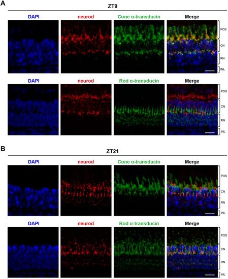
Dynamic expression of neurod in zebrafish adult photoreceptors. Representative maximum projection confocal images of double fluorescent in situ hybridisation for neurod and cone α-transducin (gnat2) or neurod and rod α-transducin (gnat1) on adult eye sections at ZT9 (A) and ZT21 (B). DAPI was used as a nuclear counterstain. POS, photoreceptor outer segments; CN, cone nuclei; RN, rod nuclei; INL, inner nuclear layer. Scale bars: 15μm. |
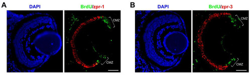
Limited photoreceptor differentiation after 5 days of zebrafish development. (A,B) Representative cross-sections of 7 dpf larval eyes (48 hours after BrdU pulse) immunostained for BrdU and the cone marker zpr-1 (A) or BrdU and the rod marker zpr-3 (B). DAPI was used as a nuclear counterstain. CMZ, circumferential marginal zone. Scale bars: 50 μm. |
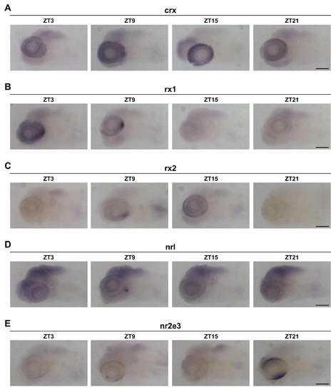
Expression of retinal transcription factors in zebrafish larvae. (A-E) Representative images of WISH for crx (A), rx1 (B), rx2 (C), nrl (D), and nr2e3 (E) on 6 dpf larvae at four different time points. Scale bars: 100 μm. |
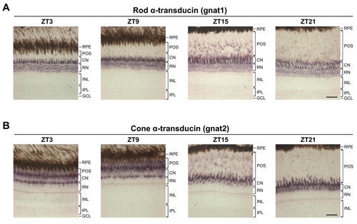
Expression of rod and cone photoreceptor markers in adult retina. (A,B) Representative images of in situ hybridisation for rod α-transducin (gnat1) (A) and cone α-transducin (gnat2) (B) on adult eye sections at four different time points. The expression pattern of each marker varies considerably throughout the day due to the positional changes of photoreceptor outer segments (retinomotor movements). Note that the gnat1 signal present in the layer of cone nuclei (CN) corresponds to rod inner segments that connect the rod nuclei (RN) to the rod outer segments. RPE, retinal pigment epithelium; POS, photoreceptor outer segments; CN, cone nuclei; RN, rod nuclei; INL, inner nuclear layer; IPL, inner plexiform layer; GCL, ganglion cell layer. Scale bars: 30 μm. |
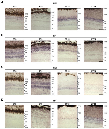
Expression of retinal transcription factors in zebrafish adult retina. (A-D) Representative images of in situ hybridisation for crx (A), rx1 (B), rx2 (C), and nrl (D) on adult eye sections at four different time points. Note that expression of nrl at ZT21 is restricted to rod photoreceptors. The signal present in the layer of cone nuclei (CN) corresponds to rod inner segments that connect the rod nuclei (RN) to the rod outer segments. RPE, retinal pigment epithelium; POS, photoreceptor outer segments; CN, cone nuclei; RN, rod nuclei; INL, inner nuclear layer; IPL, inner plexiform layer; GCL, ganglion cell layer. Scale bars: 30 μm. |
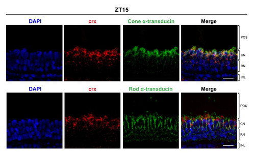
Expression of crx in zebrafish adult photoreceptors. Representative maximum projection confocal images of double fluorescent in situ hybridisation for crx and cone α-transducin (gnat2) or crx and rod α-transducin (gnat1) on adult eye sections at ZT15. DAPI was used as a nuclear counterstain. POS, photoreceptor outer segments; CN, cone nuclei; RN, rod nuclei; INL, inner nuclear layer. Scale bars: 15 μm. |