
Image
|
Figure Caption
Fig. S2
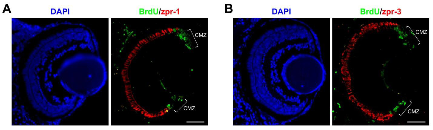
Limited photoreceptor differentiation after 5 days of zebrafish development. (A,B) Representative cross-sections of 7 dpf larval eyes (48 hours after BrdU pulse) immunostained for BrdU and the cone marker zpr-1 (A) or BrdU and the rod marker zpr-3 (B). DAPI was used as a nuclear counterstain. CMZ, circumferential marginal zone. Scale bars: 50 μm.
Acknowledgments
This image is the copyrighted work of the attributed author or publisher, and
ZFIN has permission only to display this image to its users.
Additional permissions should be obtained from the applicable author or publisher of the image.
Full text @ Development