
|
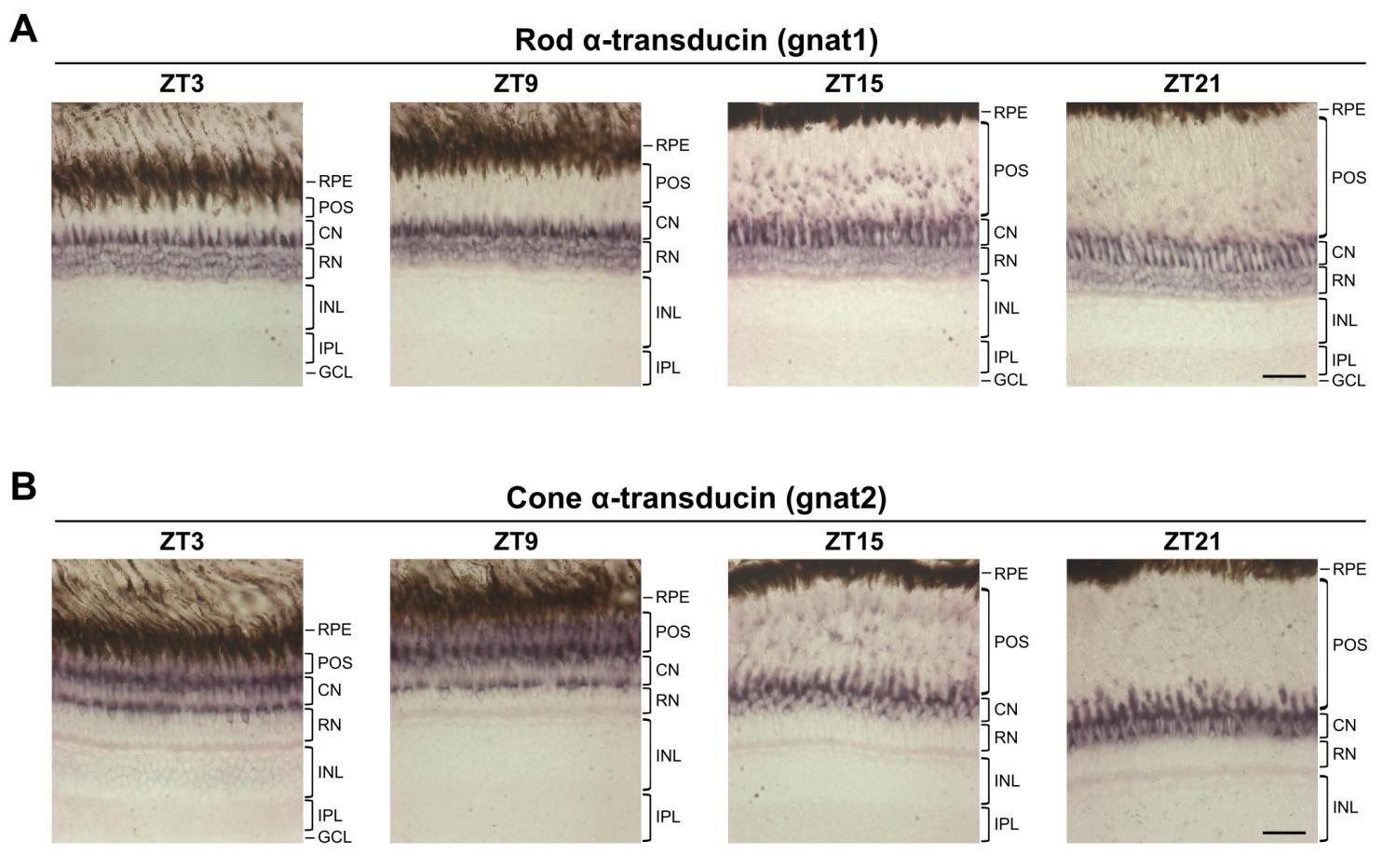
Fig. S4
Expression of rod and cone photoreceptor markers in adult retina. (A,B) Representative images of in situ hybridisation for rod α-transducin (gnat1) (A) and cone α-transducin (gnat2) (B) on adult eye sections at four different time points. The expression pattern of each marker varies considerably throughout the day due to the positional changes of photoreceptor outer segments (retinomotor movements). Note that the gnat1 signal present in the layer of cone nuclei (CN) corresponds to rod inner segments that connect the rod nuclei (RN) to the rod outer segments. RPE, retinal pigment epithelium; POS, photoreceptor outer segments; CN, cone nuclei; RN, rod nuclei; INL, inner nuclear layer; IPL, inner plexiform layer; GCL, ganglion cell layer. Scale bars: 30 μm.