
Fig. 2
- ID
- ZDB-FIG-140715-1
- Publication
- Laranjeiro et al., 2014 - Transcription factors involved in retinogenesis are co-opted by the circadian clock following photoreceptor differentiation
- Other Figures
- All Figure Page
- Back to All Figure Page
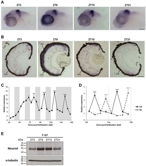
neurod rhythmic expression is restricted to retinal photoreceptors. (A,B) Representative images at four time points of neurod WISH on 6 dpf larvae (A) and of in situ hybridisation for neurod on 6 dpf larval eye sections (B). ZT, zeitgeber time; RPE, retinal pigment epithelium; ONL, outer nuclear layer. (C) qPCR analysis of neurod expression in zebrafish larvae raised on an LD cycle until 6dpf and then transferred to DD on day 7 (n=3). White and grey backgrounds represent light and dark phases, respectively. (D) qPCR analysis of neurod expression in 6-7dpf larvae raised on an LD cycle or a DL cycle (n=3). Statistically significant differences between the expression peak and trough on each day (Fisher′s LSD test) are indicated: **P<0.01, ***P<0.001. Error bars indicate s.e.m. (E) Representative western blots of Neurod and α-tubulin expression on 6dpf larvae at four time points. Scale bars: 100μm in A; 50μm in B. |