
- Title
-
Characteristics of Rod Regeneration in a Novel Zebrafish Retinal Degeneration Model Using N-Methyl-N-Nitrosourea (MNU)
- Authors
- Tappeiner, C., Balmer, J., Iglicki, M., Schuerch, K., Jazwinska, A., Enzmann, V., and Tschopp, M.
- Source
- Full text @ PLoS One

H&E staining of zebrafish retinas at baseline and after MNU exposure. A. MNU 50 mg/l group: only minor changes of retinal structure were observed after MNU treatment (e.g., disruption of the inner nuclear layer and vacuolation of the outer nuclear layer between day 1 and 30). B. MNU 150 mg/l group: Histological changes already started at day 1 (e.g., disruption of the inner nuclear layer and vacuolation of the outer nuclear layer), resulting in massive retinal degeneration with loss of nearly all rod cells at day 8. On days 8 and 15, accumulations of cell clusters (arrows) were found mainly in the inner nuclear layer. GC (ganglion cells), OPL (outer plexiform layer), INL (inner nuclear layer), RN (rod nuclei), CN (cone nuclei). Scale bar indicates 25 μm. PHENOTYPE:
|
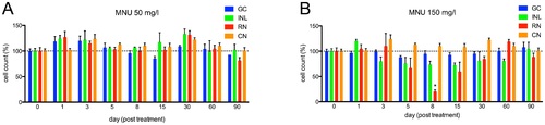
Cell counts in different retinal layers for zebrafish exposed to MNU at baseline and follow-up. A. MNU 50 mg/l group: No relevant decrease in retinal cells was observed (p>0.05). B. MNU 150 mg/l group: Rod cell loss started at day 5; the number of rods was lowest at day 8 (pd0.01) with a decrease of 79.6%, but fully recovered by day 60. Other retinal layers did not display any relevant decrease of cell numbers after MNU exposure (p>0.05). GC (ganglion cells), INL (inner nuclear layer), RN (rod nuclei) and CN (cone nuclei). Baseline values are defined as 100%. Mean values with SEM error bars are represented (* indicates pd0.01 compared with day 0). PHENOTYPE:
|
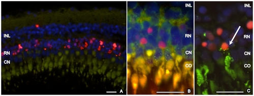
TUNEL staining of zebrafish retina, 3 days after exposure to MNU 150 mg/l. A. TUNEL-positive cells (red) are localized in the outer nuclear layer. Strong autofluorescence (green) of a cone outer segment allows identification of the corresponding nearby cone nucleus. Based on this assessment, cone photoreceptors are TUNEL-negative. B. Immunhistochemistry with zpr-1 (staining double cones, green dotted) combined with TUNEL staining (red) confirmed that cone photoreceptors are TUNEL-negative. C. Immunhistochemistry with rhodopsin (staining rods, green) and TUNEL staining (red). Co-Localization of rhodopsin and TUNEL exemplarily shows a dying rod photoreceptor (arrow). Cell nuclei are stained with DAPI (blue). INL (inner nuclear layer), RN (rod nuclei), CN (cone nuclei), CO (cone outer segment). Scale bar indicates 50 μm. PHENOTYPE:
|
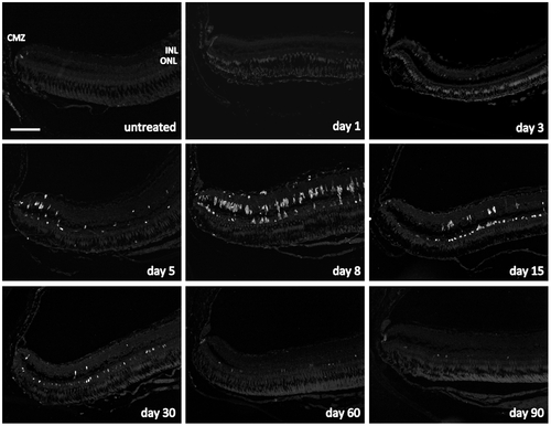
Cell proliferation in the zebrafish retina exposed to 150 mg/l MNU. Proliferating cell nuclear antigen (PCNA) positive cells (white) indicate proliferation and were found in the ciliary marginal zone (CMZ) at all time points and in the untreated fish. Furthermore, PCNA-positive cells were observed in the inner nuclear layer (INL) starting at day 3 and their number was highest at day 8. Proliferating cells in the outer nuclear layer (ONL) were not found before day 5 but their number was highest at day 15. From day 30 on, nearly no proliferating cells were seen in the INL, whereas proliferation in the ONL occurred until the end of follow up at day 90. Scale bar indicates 100 μm. PHENOTYPE:
|
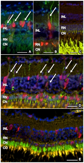
Co-localization of proliferating cells with GFAP, rhodopsin, and zpr-1. Proliferating (PCNA-positive, red) cells in the inner nuclear layer (INL), but not in the outer nuclear layer (ONL: rod nucleus = RN, cone nucleus = CN), co-localized with GFAP (green) (arrows) at days 5 (A) and 15 (B). No rhodopsin is observed in the INL of untreated retina (C), whereas many PCNA-positive cells co-localized (arrows) with rhodopsin (green) in the ONL and INL during retinal regeneration after MNU exposure (D; day 15). No co-localization of zpr-1 stained double cones (green) and PCNA (red) was found (E, day 5). Cell nuclei are stained with DAPI (blue). Scale bar indicates 50 μm. EXPRESSION / LABELING:
|