
|
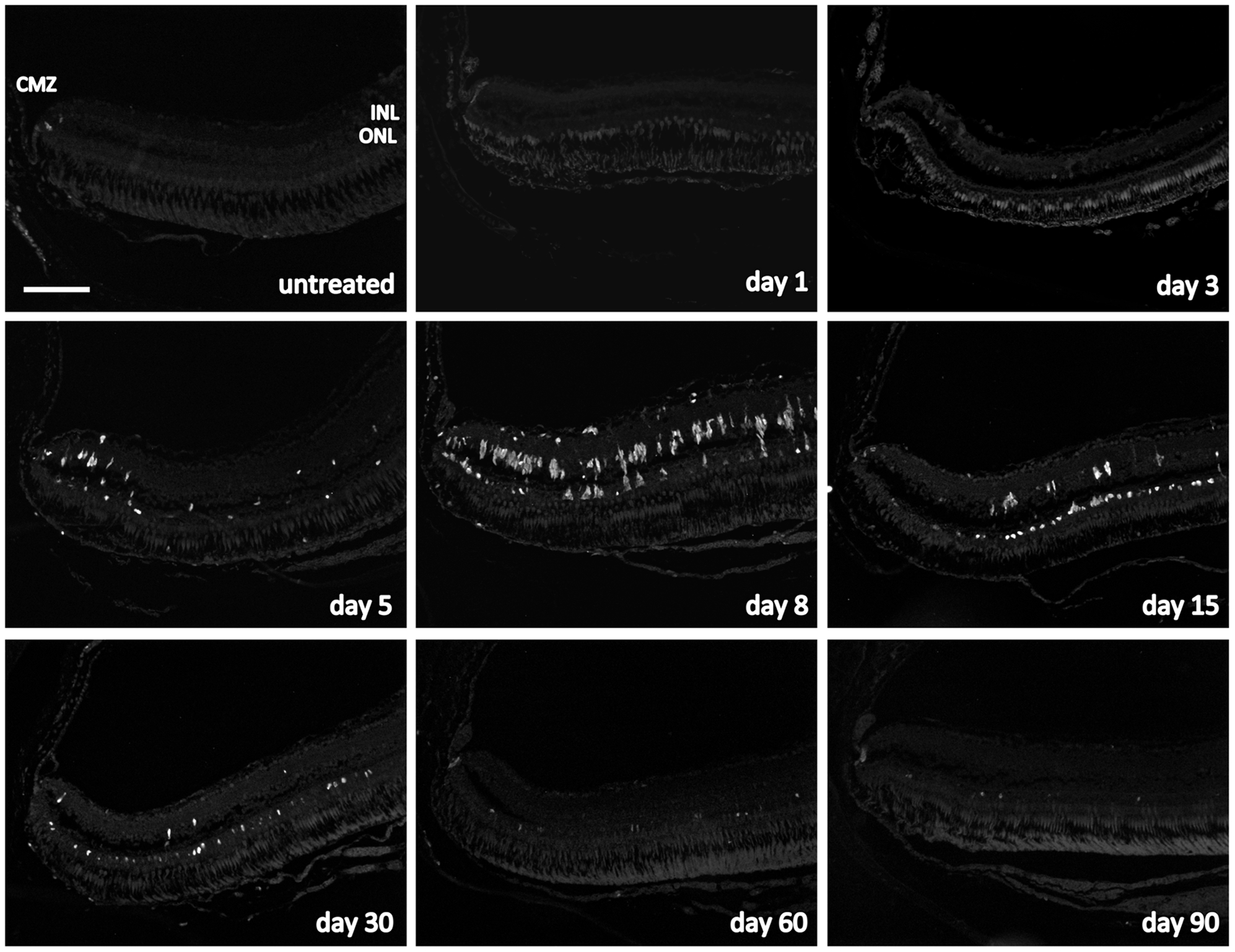
Fig. 4
Cell proliferation in the zebrafish retina exposed to 150 mg/l MNU.
Proliferating cell nuclear antigen (PCNA) positive cells (white) indicate proliferation and were found in the ciliary marginal zone (CMZ) at all time points and in the untreated fish. Furthermore, PCNA-positive cells were observed in the inner nuclear layer (INL) starting at day 3 and their number was highest at day 8. Proliferating cells in the outer nuclear layer (ONL) were not found before day 5 but their number was highest at day 15. From day 30 on, nearly no proliferating cells were seen in the INL, whereas proliferation in the ONL occurred until the end of follow up at day 90. Scale bar indicates 100 μm.