
Fig. 6
- ID
- ZDB-FIG-131015-12
- Publication
- Tappeiner et al., 2013 - Characteristics of Rod Regeneration in a Novel Zebrafish Retinal Degeneration Model Using N-Methyl-N-Nitrosourea (MNU)
- Other Figures
- All Figure Page
- Back to All Figure Page
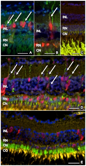
Co-localization of proliferating cells with GFAP, rhodopsin, and zpr-1. Proliferating (PCNA-positive, red) cells in the inner nuclear layer (INL), but not in the outer nuclear layer (ONL: rod nucleus = RN, cone nucleus = CN), co-localized with GFAP (green) (arrows) at days 5 (A) and 15 (B). No rhodopsin is observed in the INL of untreated retina (C), whereas many PCNA-positive cells co-localized (arrows) with rhodopsin (green) in the ONL and INL during retinal regeneration after MNU exposure (D; day 15). No co-localization of zpr-1 stained double cones (green) and PCNA (red) was found (E, day 5). Cell nuclei are stained with DAPI (blue). Scale bar indicates 50 μm. |
| Antibody: | |
|---|---|
| Fish: | |
| Condition: | |
| Anatomical Term: | |
| Stage: | Adult |