
- Title
-
Segmental assembly of fibronectin matrix requires rap1b and integrin alpha5
- Authors
- Lackner, S., Schwendinger-Schreck, J., Jülich, D., and Holley, S.A.
- Source
- Full text @ Dev. Dyn.
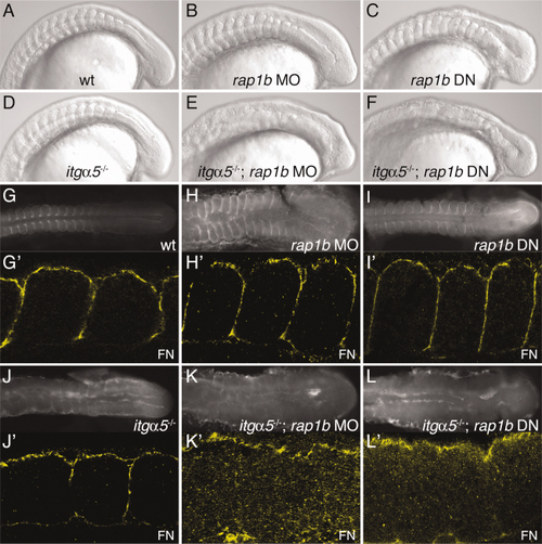
Loss of rap1b synergizes with itgα5, resulting in a disruption of all somite borders and abrogation of Fibronectin matrix assembly. A–F: Lateral view DIC images showing clear somite borders for (A) wild-type, (B) rap1b MO1-injected (8 experiments, 83.7% affected, n = 341), and (C) rap1b DN mRNA-injected (3 experiments, 100% affected, n = 177) embryos. For synergy experiments, rap1b MO1 or rap1b DN was injected into the progeny of heterozygous itgα5+/- crosses. Therefore, 100% penetrance would result in approximately 25% of embryos exhibiting the phenotype. D: Anterior somite borders are disrupted in the itgα5-/- mutant, while (E) itgα5-/-; rap1b MO1 (9 experiments, 27.4% affected, n = 865) and (F) itgα5-/-; rap1b DN (6 experiments, 22.3% affected, n = 455) embryos lack somite borders along the entire AP axis. (G- L) Dorsal view and (G′-L′) higher magnification dorsal view of FN localization showing clear FN matrix surrounding somites in (G–G′) wild-type, (H–H′) rap1b MO1, and (I–I′) rap1b DN embryos. J,J′: FN matrix surrounds only posterior somites in itgα5-/- mutants, but few fibrils are seen within the paraxial mesoderm of either (K,K′) itgα5-/-; rap1b MO1 or (L,L′) itgα5-/-; rap1b DN embryos. For all images, anterior is to the left and embryos are at the 12–15-somite stage. EXPRESSION / LABELING:
PHENOTYPE:
|
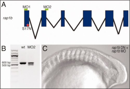
Morpholino knockdown of rap1b is specific. A: Schematic of the rap1b gene showing exons in blue, introns in black, and the approximate position of morpholino binding sites in green. The S17N mutation generating dominant-negative rap1b is indicated with a red star. B: Agarose gel showing 100-bp ladder with 500- and 600-bp bands marked, PCR product of wild-type rap1b cDNA, and PCR product of wild-type and altered splice product generated via splice-blocking morpholino (MO2) for rap1b. As confirmed by sequencing, the altered splice-product lacks the second exon and is 69 bp smaller than the wild-type rap1b cDNA. This disruption of splicing results in deletion of 23 amino acids from the predicted protein. C: Lateral view of DIC image for 12–15-somite-stage embryo injected with both rap1b MO1 and rap1b DN mRNA (3 experiments). For rap1b MO1+MO2 injections, 71.4% were affected (3 experiments, n = 248) (data not shown). Anterior is left. |
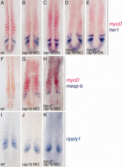
Loss of somite borders is likely not due to patterning defects in the PSM. A-E: Double in situ hybridization for myoD (pink) and her1 (blue) for (A) wild-type, (B) rap1b MO1, (C) rap1b DN mRNA, (D) itgα5-/-; rap1b MO1, and (E) itgα5-/-; rap1b DN 12-15-somite-stage embryos. Stripes of her1 expression persist in all backgrounds. In contrast, segmental myoD expression is blurred in rap1b MO1 and rap1b DN embryos, and completely lost in itgα5-/-; rap1b MO or itgα5-/-; rap1b DN. F-H: Double in situ hybridization for myoD (pink) and mesp-b (blue) in (F) wild-type, (G) rap1b MO1, and (H) itgα5-/-; rap1b MO1 embryos at 12-15-somite stage. Stripes of mesp-b can be seen in both rap1b and itgα5-/-; rap1b morphants. I-K: In situ hybridization for ripply1 in (I) wild-type, (J) rap1b MO1, and (K) itgα5-/-; rap1b MO1 12-15-somite stage embryos. Segmental ripply1 expression is disrupted only in itgα5-/-; rap1b MO1 embryos. For all images, anterior is up. EXPRESSION / LABELING:
|
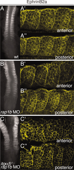
Localization of EphrinB2a suggests gradual loss of segment polarity after loss of both rap1b and itg±5 function. Dorsal views (A–C:) and higher magnification dorsal views (A’–C’’) of EphrinB2a protein localized to the posterior halves of somites in (A–A’’) wild-type and (B–B’’) rap1b MO1 embryos. Localized EphrinB2a is lost in anterior somites of (C–C’’) itg±5/; rap1b double morphants, but is transiently present in more recently formed posterior somites. Shown are 12–15-somite-stage zebrafish embryos. In A–C, anterior is up, while in A’–C’’ anterior is left. |
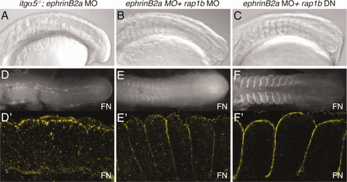
Somite morphogenesis is sensitive to genetic interaction between itgα5 and ephrinB2a, but not rap1b and ephrinB2a. A-C: Lateral view DIC images showing complete disruption of somite borders in (A) itgα5-/-; ephrinB2a MO embryos (7 experiments, 27.9% affected compared to an expected 25% for homozygotes, n = 605) and proper maintenance of somite borders in (B) ephrinB2a MO + rap1b MO1 (5 experiments, no synergy, n = 455) and (C) ephrinB2a MO + rap1b DN embryos (3 experiments, no synergy). Dorsal (D-F:) and higher magnification dorsal (D′-F′) views of Fibronectin localization for (D-D′) itgα5-/-; ephrinB2a MO, (E-E′) ephrinB2a MO + rap1b MO1, and (F-F′) ephrinB2a MO + rap1b DN embryos at the 12–15-somite stage. FN matrix formation is lost only in itgα5-/-; ephrinB2a MO embryos. For all images, anterior is left. EXPRESSION / LABELING:
PHENOTYPE:
|