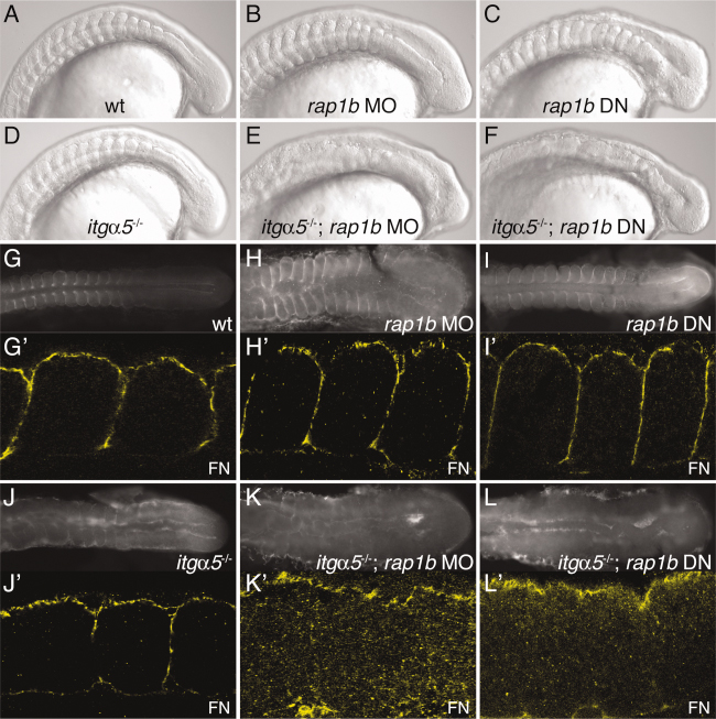
Fig. 1 Loss of rap1b synergizes with itgα5, resulting in a disruption of all somite borders and abrogation of Fibronectin matrix assembly. A–F: Lateral view DIC images showing clear somite borders for (A) wild-type, (B) rap1b MO1-injected (8 experiments, 83.7% affected, n = 341), and (C) rap1b DN mRNA-injected (3 experiments, 100% affected, n = 177) embryos. For synergy experiments, rap1b MO1 or rap1b DN was injected into the progeny of heterozygous itgα5+/- crosses. Therefore, 100% penetrance would result in approximately 25% of embryos exhibiting the phenotype. D: Anterior somite borders are disrupted in the itgα5-/- mutant, while (E) itgα5-/-; rap1b MO1 (9 experiments, 27.4% affected, n = 865) and (F) itgα5-/-; rap1b DN (6 experiments, 22.3% affected, n = 455) embryos lack somite borders along the entire AP axis. (G- L) Dorsal view and (G′-L′) higher magnification dorsal view of FN localization showing clear FN matrix surrounding somites in (G–G′) wild-type, (H–H′) rap1b MO1, and (I–I′) rap1b DN embryos. J,J′: FN matrix surrounds only posterior somites in itgα5-/- mutants, but few fibrils are seen within the paraxial mesoderm of either (K,K′) itgα5-/-; rap1b MO1 or (L,L′) itgα5-/-; rap1b DN embryos. For all images, anterior is to the left and embryos are at the 12–15-somite stage.
Image
Figure Caption
Figure Data
Acknowledgments
This image is the copyrighted work of the attributed author or publisher, and
ZFIN has permission only to display this image to its users.
Additional permissions should be obtained from the applicable author or publisher of the image.
Full text @ Dev. Dyn.

