
|
Fig. 3
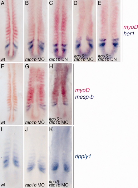
Loss of somite borders is likely not due to patterning defects in the PSM. A-E: Double in situ hybridization for myoD (pink) and her1 (blue) for (A) wild-type, (B) rap1b MO1, (C) rap1b DN mRNA, (D) itgα5-/-; rap1b MO1, and (E) itgα5-/-; rap1b DN 12-15-somite-stage embryos. Stripes of her1 expression persist in all backgrounds. In contrast, segmental myoD expression is blurred in rap1b MO1 and rap1b DN embryos, and completely lost in itgα5-/-; rap1b MO or itgα5-/-; rap1b DN. F-H: Double in situ hybridization for myoD (pink) and mesp-b (blue) in (F) wild-type, (G) rap1b MO1, and (H) itgα5-/-; rap1b MO1 embryos at 12-15-somite stage. Stripes of mesp-b can be seen in both rap1b and itgα5-/-; rap1b morphants. I-K: In situ hybridization for ripply1 in (I) wild-type, (J) rap1b MO1, and (K) itgα5-/-; rap1b MO1 12-15-somite stage embryos. Segmental ripply1 expression is disrupted only in itgα5-/-; rap1b MO1 embryos. For all images, anterior is up.