
- Title
-
Evidence of a role of inositol polyphosphate 5-phosphatase INPP5E in cilia formation in zebrafish
- Authors
- Luo, N., Lu, J., and Sun, Y.
- Source
- Full text @ Vision Res.
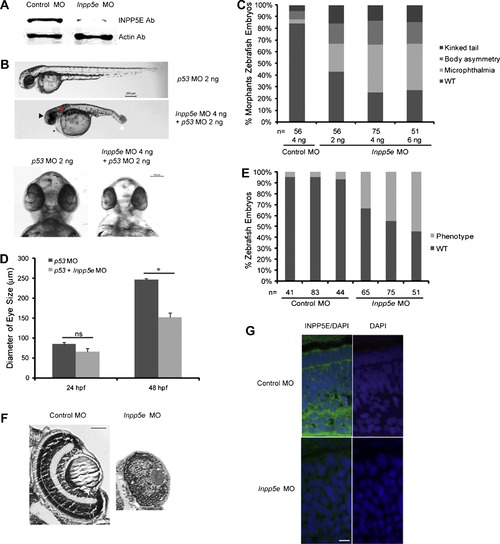
Phenotype of Inpp5e morphants. (A) Immunoblot analysis of 30 µg of total lysates of zebrafish embryo injected with control MO (4 ng) or Inpp5e MO (4 ng) at 48 hpf with anti-INPP5E and anti-²-actin antibodies. (B) Zebrafish embryos were injected with p53 MO (2 ng) or p53 MO (2 ng) and Inpp5e MO (4 ng). Representative phenotypes of microphthalmia (black arrowhead), pericardial edema (small arrow), body axis asymmetry, kinked tail (white arrow), pronephric cyst formation (red arrow), and hypopigmentation were observed at 48 hpf (top panel, scale bar 250 µm). The ventral sides of morphants are shown (bottom, scale bar 100 µm). (C) Dose-dependent effect of morpholinos in zebrafish. Control and Inpp5e MO at indicated doses were injected into zebrafish embryos, and phenotypes of microphthalmia, kinked tail, and body asymmetry were quantified at 48 hpf (ANOVA, F = 92, p = 3.6E10), kinked tail (ANOVA, F = 3.6, p = 0.08), and body asymmetry (ANOVA, F = 5.2, p = 0.04) (n = the number of injected embryos). (D) Quantification of eye size of morphants at 24 hpf and 48 hpf. The eye size was determined by the longest diameters of eye balls in dorsal view (n = 20 embryos, three independent experiments, unpaired t-test, * p = 1.2E08, ns means not statistically significant). (E) Inpp5e MO1 (4 ng) and control MO (4 ng) were injected into 1-cell zebrafish embryos. At 48 hpf, all the phenotypes were assessed and the total numbers of defective morphants in four independent experiments were quantified (n, the number of injected embryos, unpaired t-test, p = 0.02). (F) Cresyl violet staining of ocular sections of zebrafish larvae (5 dpf) injected with control MO (4 ng) or Inpp5e MO (4 ng). Scale bar 30 µm. (G) Immunostaining of 3 dpf zebrafish larvae injected with control MO (4 ng) or Inpp5e MO (4 ng), followed by antibody staining against INPP5E (green) (DAPI, blue). Scale bar 10 µm. EXPRESSION / LABELING:
|
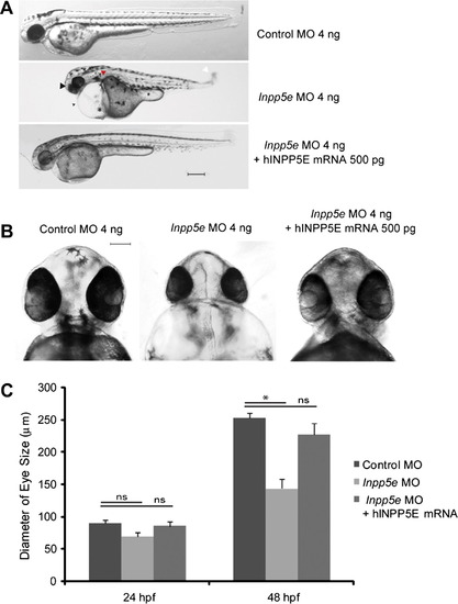
Human INPP5E rescue phenotypes of Inpp5e morphants. (A) Zebrafish embryos were injected with control MO (4 ng), Inpp5e MO (4 ng) or Inpp5e MO (4 ng) and human INPP5E mRNA (hINPP5E mRNA, 500 pg). Representative phenotypes of microphthalmia (arrowhead), pericardial edema (arrow), body axis asymmetry, kinked tail (white arrow), pronephric cyst formation (red arrow), and hypopigmentation were observed at 48 hpf (scale bar 250 µm). (B) The ventral sides of embryos were injected with control MO (4 ng), Inpp5e MO (4 ng) or Inpp5e MO (4 ng) and human INPP5E mRNA (hINPP5E mRNA, 500 pg), scale bar 100 µm. (C) Quantification of eye size of zebrafish morphants at 24 hpf and 48 hpf (N = 20 embryos, three independent experiments, unpaired t-test, * p = 1.61E12). |
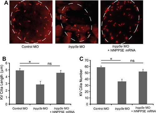
Kupffer’s vesicle cilia defect in Inpp5e morphants. (A) Human INPP5E mRNA could rescue the loss of Inpp5e. KV cilia of zebrafish embryos injected with control MO (4 ng), Inpp5e MO (4 ng) or Inpp5e MO (4 ng) and hINPP5E mRNA (500 ng) at 6-somite stage were immunostained with acetylated α-tubulin (red), representative images are shown (dash line indicates border of KV). (B and C) Quantification of number (B) and length (C) of KV cilia in zebrafish embryos injected with control MO (4 ng), Inpp5e MO (4 ng) or Inpp5e MO (4 ng) and hINPP5E mRNA (500 ng) (N = 20 embryos, three independent experiments, unpaired t-test, * p = 0.02 in (B) and * p = 1.56E08 in (C)). PHENOTYPE:
|
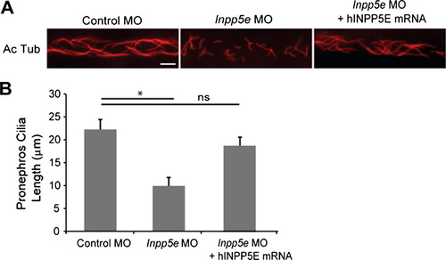
Pronephric cilia defect in Inpp5e morphants. (A) INPP5E mRNA rescue of Inpp5e pronephric cilia formation. Representative image of pronephric cilia of zebrafish embryos at 24 hpf stage, injected with control MO (4 ng), Inpp5e MO (4 ng) or Inpp5e MO (4 ng) and hINPP5E mRNA (500 ng), immunostaining with acetylated α-tubulin (red). Scale bar 10 µm. (B) Pronephric cilia length of control and Inpp5e MO. Pronephric cilia of zebrafish embryos injected with control MO (4 ng), Inpp5e MO (4 ng) or Inpp5e MO (4 ng) and hINPP5E mRNA (500 ng) at 24 hpf stage were analyzed by immunostaining with acetylated α-tubulin and cilia length was measured (N = 20 embryos, three independent experiments, unpaired t-test, * p = 8.57E08). PHENOTYPE:
|
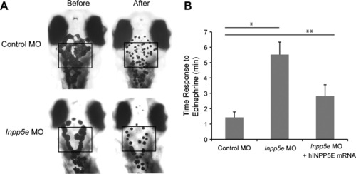
Impaired melanosome transport in Inpp5e morphants. (A) Inpp5e morphants showed slowed retrograde melanosome transport. Five day-old larvas injected with morpholinos were treated with epinephrine, and the time required for all melanosomes in the head and trunk to retract was determined. Representative photos are shown. (B) Quantification of the response time for epinephrine treatments in the control MO (2 ng), Inpp5e MO (2 ng), and Inpp5e MO (2 ng) and hINPP5E mRNA (400 pg) embryos (N = 20 embryos, three independent experiments, unpaired t-test, * p = 1.17E18, ** p = 1.5E12). PHENOTYPE:
|
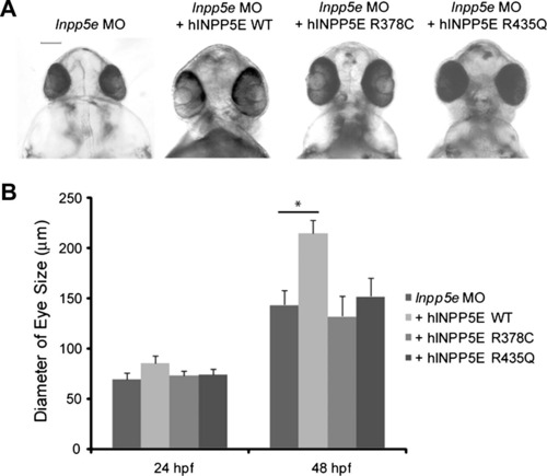
INPP5E mutants in zebrafish eye development. (A) Zebrafish embryos were injected with Inpp5e MO (4 ng), Inpp5e MO (4 ng) and hINPP5E WT mRNA (500 pg), Inpp5e MO (4 ng) and hINPP5E R378C mRNA (500 pg), or Inpp5e MO (4 ng) and hINPP5E R435Q mRNA (500 pg). The ventral sides of morphants are shown, scale bar 100 µm. (B) Quantification of eye size of morphants at 24 hpf and 48 hpf (N = 20 embryos, three independent experiments, unpaired t-test, * p = 3.6E08). |
Reprinted from Vision Research, 75, Luo, N., Lu, J., and Sun, Y., Evidence of a role of inositol polyphosphate 5-phosphatase INPP5E in cilia formation in zebrafish, 98-107, Copyright (2012) with permission from Elsevier. Full text @ Vision Res.