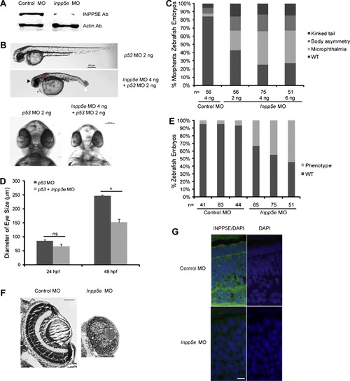
Phenotype of Inpp5e morphants. (A) Immunoblot analysis of 30 µg of total lysates of zebrafish embryo injected with control MO (4 ng) or Inpp5e MO (4 ng) at 48 hpf with anti-INPP5E and anti-²-actin antibodies. (B) Zebrafish embryos were injected with p53 MO (2 ng) or p53 MO (2 ng) and Inpp5e MO (4 ng). Representative phenotypes of microphthalmia (black arrowhead), pericardial edema (small arrow), body axis asymmetry, kinked tail (white arrow), pronephric cyst formation (red arrow), and hypopigmentation were observed at 48 hpf (top panel, scale bar 250 µm). The ventral sides of morphants are shown (bottom, scale bar 100 µm). (C) Dose-dependent effect of morpholinos in zebrafish. Control and Inpp5e MO at indicated doses were injected into zebrafish embryos, and phenotypes of microphthalmia, kinked tail, and body asymmetry were quantified at 48 hpf (ANOVA, F = 92, p = 3.6E10), kinked tail (ANOVA, F = 3.6, p = 0.08), and body asymmetry (ANOVA, F = 5.2, p = 0.04) (n = the number of injected embryos). (D) Quantification of eye size of morphants at 24 hpf and 48 hpf. The eye size was determined by the longest diameters of eye balls in dorsal view (n = 20 embryos, three independent experiments, unpaired t-test, * p = 1.2E08, ns means not statistically significant). (E) Inpp5e MO1 (4 ng) and control MO (4 ng) were injected into 1-cell zebrafish embryos. At 48 hpf, all the phenotypes were assessed and the total numbers of defective morphants in four independent experiments were quantified (n, the number of injected embryos, unpaired t-test, p = 0.02). (F) Cresyl violet staining of ocular sections of zebrafish larvae (5 dpf) injected with control MO (4 ng) or Inpp5e MO (4 ng). Scale bar 30 µm. (G) Immunostaining of 3 dpf zebrafish larvae injected with control MO (4 ng) or Inpp5e MO (4 ng), followed by antibody staining against INPP5E (green) (DAPI, blue). Scale bar 10 µm.
|
