
Fig. 4
- ID
- ZDB-FIG-130129-4
- Publication
- Luo et al., 2012 - Evidence of a role of inositol polyphosphate 5-phosphatase INPP5E in cilia formation in zebrafish
- Other Figures
- All Figure Page
- Back to All Figure Page
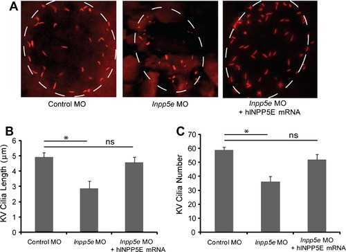
Kupffer’s vesicle cilia defect in Inpp5e morphants. (A) Human INPP5E mRNA could rescue the loss of Inpp5e. KV cilia of zebrafish embryos injected with control MO (4 ng), Inpp5e MO (4 ng) or Inpp5e MO (4 ng) and hINPP5E mRNA (500 ng) at 6-somite stage were immunostained with acetylated α-tubulin (red), representative images are shown (dash line indicates border of KV). (B and C) Quantification of number (B) and length (C) of KV cilia in zebrafish embryos injected with control MO (4 ng), Inpp5e MO (4 ng) or Inpp5e MO (4 ng) and hINPP5E mRNA (500 ng) (N = 20 embryos, three independent experiments, unpaired t-test, * p = 0.02 in (B) and * p = 1.56E08 in (C)). |
| Fish: | |
|---|---|
| Knockdown Reagent: | |
| Observed In: | |
| Stage: | 5-9 somites |
Reprinted from Vision Research, 75, Luo, N., Lu, J., and Sun, Y., Evidence of a role of inositol polyphosphate 5-phosphatase INPP5E in cilia formation in zebrafish, 98-107, Copyright (2012) with permission from Elsevier. Full text @ Vision Res.