
Fig. 5
- ID
- ZDB-FIG-130129-5
- Publication
- Luo et al., 2012 - Evidence of a role of inositol polyphosphate 5-phosphatase INPP5E in cilia formation in zebrafish
- Other Figures
- All Figure Page
- Back to All Figure Page
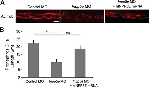
Pronephric cilia defect in Inpp5e morphants. (A) INPP5E mRNA rescue of Inpp5e pronephric cilia formation. Representative image of pronephric cilia of zebrafish embryos at 24 hpf stage, injected with control MO (4 ng), Inpp5e MO (4 ng) or Inpp5e MO (4 ng) and hINPP5E mRNA (500 ng), immunostaining with acetylated α-tubulin (red). Scale bar 10 µm. (B) Pronephric cilia length of control and Inpp5e MO. Pronephric cilia of zebrafish embryos injected with control MO (4 ng), Inpp5e MO (4 ng) or Inpp5e MO (4 ng) and hINPP5E mRNA (500 ng) at 24 hpf stage were analyzed by immunostaining with acetylated α-tubulin and cilia length was measured (N = 20 embryos, three independent experiments, unpaired t-test, * p = 8.57E08). |
| Fish: | |
|---|---|
| Knockdown Reagent: | |
| Observed In: | |
| Stage: | Prim-5 |
Reprinted from Vision Research, 75, Luo, N., Lu, J., and Sun, Y., Evidence of a role of inositol polyphosphate 5-phosphatase INPP5E in cilia formation in zebrafish, 98-107, Copyright (2012) with permission from Elsevier. Full text @ Vision Res.