
- Title
-
Dually inducible TetON systems for tissue-specific conditional gene expression in zebrafish
- Authors
- Knopf, F., Schnabel, K., Haase, C., Pfeifer, K., Anastassiadis, K., and Weidinger, G.
- Source
- Full text @ Proc. Natl. Acad. Sci. USA
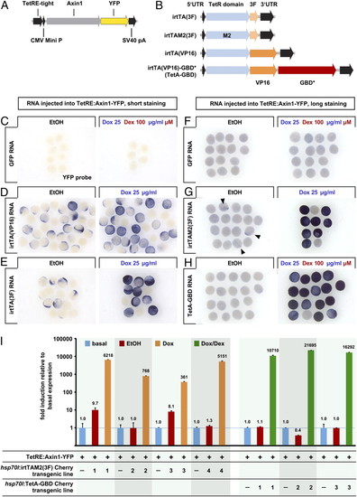
Dually inducible TetA-GBD activates transgene transcription in a nonleaky fashion in zebrafish embryos. (A) Transgenic Tet responder construct. (B) Human codon optimized (“improved”) variants of the reverse tetracycline-responsive transactivator (irtTA) used in this study. (C–H) YFP RNA expression detected by whole-mount in situ hybridization in TetRE:Axin1-YFP transgenic embryos injected with equimolar amounts of GFP (25 pg), irtTA(VP16) (30 pg), irtTA(3F) (25 pg), irtTAM2(3F) (30 pg), or irtTA(VP16)-GBD* (TetA-GBD, 50 pg) and treated with EtOH vehicle or 25 μg/mL Dox or 25 μg/mL Dox plus 100 μM Dex from 5 hpf for 4.5h (C–E) or 3.5h (F–H). Samples in F–H were stained significantly longer than those in C–E to reveal even low levels of leakiness. Note severe leaky induction in irtTA(VP16) and irtTA(3F) injected embryos and weak leakiness in irtM2(3F) injected embryos (arrowheads). (I) Axin1-YFP RNA expression detected by QPCR in progeny of TetRE:Axin1-YFP fish crossed with individual sublines of hsp70l:irtTAM2(3F)-p2a-mCherry or hsp70l:TetA-GBD-p2a-mCherry transgenic fish, heatshocked at 24 hpf, and treated with EtOH or 25 μg/mL Dox or Dox plus 100 μM Dex for 4 h. Levels are normalized to expression in TetRE:Axin1-YFP embryos containing no TetActivator transgene (“basal”). |
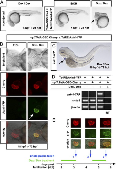
Tissue-specific, reversible gene expression using the dually inducible TetA-GBD/TetRE-tight system in embryonic and adult zebrafish. (A) Severe Wnt/β-catenin loss-of-function phenotypes as evidenced by posterior truncations and expanded eyes (arrow) in TetRE:Axin1-YFP embryos injected with 100 ng/μl TetA-GBD RNA and treated with Dox/Dex from 4 h postfertilization (hpf) until 24 hpf. Note that embryos treated with EtOH vehicle and noninjected embryos treated with Dox/Dex develop normally. n = 30 noninj, 7 EtOH, 9 Dox/Dex. (B) Induction of Axin1-YFP expression in ventricle (arrow) in myl7:TetA-GBD Cherry; TetRE:Axin1-YFP double transgenic embryos treated with Dox/Dex for 24 h from 48 hpf. n = 15 EtOH, 18 Dox/Dex. (C) Heart-specific induction of Axin1-YFP RNA (arrow) after Dox/Dex treatment. n = 15. (D) Semiquantitative PCR detects axin1-YFP expression only in myl7:TetA-GBD Cherry; TetRE:Axin1-YFP double transgenic embryos treated with Dox/Dex (lane 3), but not in EtOH treated embryos (lane 2) or embryos only containing the TetRE:Axin1-YFP transgene (lane 1). myl7 and β-actin are shown as loading controls. (E) Fast reversibility and reinducibility of Axin1-YFP induction. Fluorescent images of the heart of one individual myl7:TetA-GBD; TetRE:Axin1-YFP double transgenic embryo is shown that was treated with Dox/Dex and photographed at the times indicated. Note that YFP signal is not detectable 24 h after drug withdrawal (Middle, column 2) and reexpressed after additional 24 h of drug treatment (Middle, column 3). n = 5. |
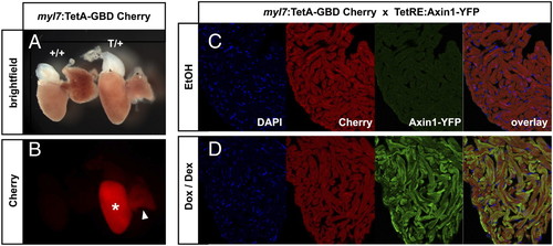
(A and B) The myl7:TetA-GBD Cherry transgene is expressed in the adult heart. (A) Brightfield images of whole extracted adult hearts: (Left) WT; (Right) myl7:TetA-GBD Cherry heterozygous. (B) Cherry channel. Note Cherry expression in ventricle (*) and weakly in atrium (arrowhead). (C and D) Inducibility in adult hearts. Cryosections of adult hearts of myl7:TetA-GBD Cherry; TetRE:Axin1-YFP double transgenic fish that had been injected intraperitoneally with EtOH (C) or 20 μg Dox plus 5 μg Dex (D). Axin1-YFP is detected by anti-GFP antibody. n = 6 EtOH and Dox/Dex. |
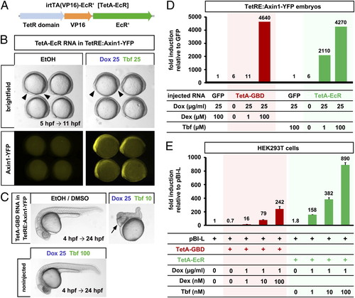
An ecdysone receptor-based dually inducible TetActivator. (A) TetA-EcR construct. (B) Induction of Axin1-YFP in TetRE:Axin1-YFP transgenic embryos injected with 100 pg of TetA-EcR RNA, treated with 25 μg/mL Dox and 25 μM Tebufenozide (Tbf) from 5 hpf and photographed at 11 hpf. Note shortened body axes (arrowheads) indicative of Axin1 gain-of-function phenotypes in drug-treated embryos. n = 10/group. (C) Severe Axin1 overexpression phenotype (posterior truncations and expanded anterior fates; arrow) in 25 μg/mL Dox and 10 μM Tbf drug-treated embryos at 24 hpf. Note that embryos treated with EtOH/DMSO vehicle and noninjected embryos treated with even higher doses of Dox/Tbf develop normally. n = 32 noninj, 6 EtOH/DMSO, 6 Dox/Tbf. (D) TetA-EcR induces more strongly than TetA-GBD at low drug doses in zebrafish. TetRE:Axin1-YFP embryos were injected with equimolar amounts of GFP (25 pg), TetA-GBD (50 pg) or TetA-EcR (55 pg) RNA, treated with the indicated drugs at 5 hpf for 3 h, YFP expression was quantified using QPCR and is shown relative to GFP controls treated with the same drugs. Zero indicates EtOH vehicle only. (E) TetA-EcR is more efficient than TetA-GBD in mammalian cells. HEK293 cells were transiently transfected with the Tet responsive luciferase reporter pBI-L and the activators TetA-GBD or TetA-EcR, and luciferase levels were measured after treatment with indicated doses of drugs. Levels are shown relative to cells transfected with pBI-L only. Error bars represent SEM. |
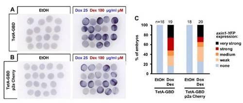
TetA-GBD tagged with p2a Cherry is functional. (A and B) YFP RNA expression detected by whole-mount in situ hybridization in TetRE:Axin1-YFP transgenic embryos injected with equimolar amounts of TetA-GBD (50 pg) or TetA-GBD p2a Cherry (65 pg), and treated with EtOH vehicle or 25 μg/mL Dox plus 100 μM Dex from 5 h postfertilization (hpf) for 3.5 h. (C) Quantification of data presented in A and B. Axin1-YFP RNA expression levels were classified into five classes, and the number of embryos in each class was counted. |
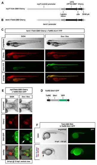
Conditional, spatially controlled induction using additional TetA-GBD drivers and TetRE responders. (A) Transgenic driver construct for expression of TetA-GBD Cherry specifically in the myocardium. (B) Transgenic driver construct for expression of TetA-GBD Cherry specifically in her4.1 expression domains, primarily in the CNS. (C) her4.1:TetA-GBD Cherry drives efficient induction of Axin1-YFP in embryos doubly transgenic with TetRE:Axin1-YFP. Embryos were treated with EtOH or 25 μg/mL Dox plus 100 μM Dex from 24 h postfertilization (hpf) and photographed at 48 hpf. (D) Transgenic responder line construct for expression of Dkk1-GFP. (E) Heart-specific expression of Dkk1-GFP in myl7:TetA-GBD Cherry; TetRE:Dkk1-GFP double transgenic fish. Images of double transgenic embryos at 72 hpf, treated with EtOH vehicle or 25 μg/mL Dox plus 100 μM Dex from 48 hpf. Note that secreted Dkk1-GFP protein accumulates in the pericardial sac (arrow) in Dox/Dex-treated embryos. *Yolk background fluorescence. n = 20 EtOH, 24 Dox/Dex. (F) Wnt/β-catenin loss-of-function phenotypes as evidenced by posterior truncations and expanded eyes (arrow) in TetRE:Dkk1-GFP embryos injected with 95 pg TetA-GBD RNA and treated with 10 μg/mL Dox plus 100 μM Dex from 4 hpf until 24 hpf. n = 7 EtOH, 8 Dox/Dex. |
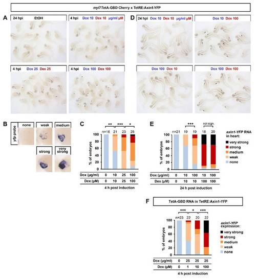
Drug dose dependency of Axin1-YFP induction in myl7:TetA-GBD Cherry; TetRE:Axin1-YFP double transgenic embryos. (A) Dose–response to indicated doses of drugs applied at 48 h postfertilization (hpf). YFP RNA was detected by whole-mount in situ hybridization 4 h postinduction, EtOH control-treated embryos are shown at 24 h postinduction. (B) Classes of YFP expression in heart used for quantification. (C) Quantification of data shown in A. **P = 0.002 (Fisher exact test), ***P < 0.001 (χ2 test), *P = 0.034 (χ2 test). (D) Dex is limiting for induction in myl7:TetA-GBD Cherry; TetRE:Axin1-YFP double transgenic embryos. The indicated doses of drugs were applied at 48 hpf, YFP RNA was detected by whole-mount in situ hybridization 24 h postinduction. (E) Quantification of data shown in D). ***P < 0.001 (χ2 test). (F) Dex is limiting for induction in TetRE:Axin1-YFP embryos injected with 90 pg TetA-GBD RNA. Embryos were treated with indicated doses of drugs at 7 hpf for 4.5 h. Axin1-YFP expression was detected by in situ hybridization and classified as in Fig S1. ***P < 0.001, *P = 0.019 (χ2 test). |
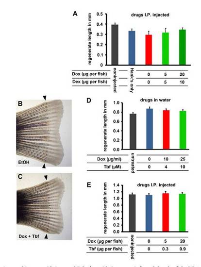
Combined treatment with Dox and Dex or with Dox and Tebufenozide is not toxic for adult zebrafish. (A) Average regenerate length at 8 d postamputation (dpa) of fish intraperitoneally injected with the indicated doses of drugs at 1 dpa, 3 dpa, and 6 dpa. Zero indicates EtOH vehicle only. Regenerate length is not significantly different (Student t test). Error bars indicate SEM. n = 6/group. (B and C) Representative regenerating fins at 7 dpa of fish housed in water containing vehicle or 25 μg/mL Dox plus 10 μM Tebufenozide. Note robust regeneration in both. Amputation plane is indicated by arrowheads. (D) Average regenerate length measured at 7 dpa of fish incubated with the indicated doses of drugs from 0.5 dpa. Zero indicates vehicle only. Regenerate length is not significantly different between vehicle control and drug-treated fins (Student t test). Error bars are SEM. n = 6/Group. (E) Average regenerate length at 8 dpa of fish intraperitoneally injected with the indicated doses of drugs at 1 dpa, 3 dpa, and 6 dpa. Zero indicates vehicle only. Regenerate length is not significantly different (Student t test). Error bars indicate SEM. n = 6/Group. |