
- Title
-
Phenotypic analysis of images of zebrafish treated with Alzheimer's gamma-secretase inhibitors
- Authors
- Arslanova, D., Yang, T., Xu, X., Wong, S.T., Augelli-Szafran, C.E., and Xia, W.
- Source
- Full text @ BMC Biotechnol.
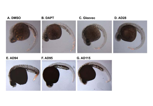
Apoptosis in zebrafish embryos treated with compounds. Zebrafish embryos were treated with GSI or fragments of Gleevec at 6 hpf. Embryos were then fixed and subjected to TUNEL staining at 24 hpf. Control embryos were treated with 0.1% DMSO and the remaining embryos were treated with different GSIs or Gleevec fragments at 50 μM, except for AD115 whose concentration was 10 μM. The majority of apoptotic cells were observed in the fish tail, but no difference among individual compound-treated embryos was observed. The representative images from 2 different experiments are presented. |
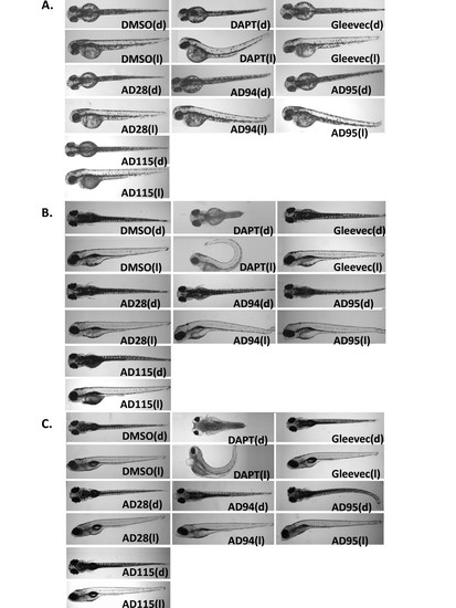
Phenotypes of the embryos treated with GSI, Gleevec, and fragments at 24 hpf. Dorsal view (indicated as "d", top panel) and lateral view (indicated as "l", bottom panel) of embryos are shown. Control embryos treated with 0.1% DMSO show a wild-type phenotype. Embryos treated with GSI, DAPT, resulted in defects in somitogenesis, eye development, pigmentation, and trunk formation as observed at 2 dpf (A), 4 dpf (B), and 6 dpf (C). Embryos treated with DAPT, AD94 or AD95 started to show defects at 2 dpf, and phenotypes of remaining embryos were most obvious at 4 or 6 dpf. The following concentrations were used: 50 μM DAPT, 50 μM Gleevec, 50 μM AD28, 50 μM AD94, 50 μM AD95, and 10 μM AD115. |
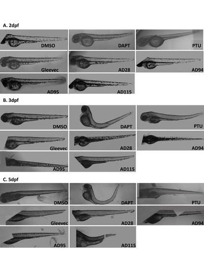
Automatically acquired images of embryos treated with GSI or fragments of Gleevec during early development stage (6 hpf). Embryos were treated with GSIs or Gleevec fragments at the following concentrations at 6 hpf: 0.1%DMSO, 50 μM for all compounds, except for 10 μM AD115. Images of living zebrafish in microtiter plates were acquired by In Cell Analyzer at 2 dpf (A), 3 dpf (B) and 5 dpf (C), and several fields of images from the individual wells were automatically merged. Because the instrument acquired several fields that covered a portion of the microtiter plate well, some images show truncations of the zebrafish. |
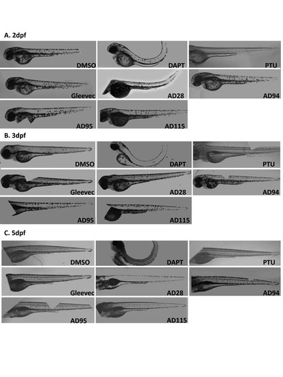
Automatically acquired images of embryos treated with GSI or fragments of Gleevec at 24 hpf. Embryos were treated with GSIs and fragments of Gleevec at 24 hpf, and 50 μM of all compounds, except for 10 μM of AD115, were applied to these embryos. Images of living zebrafish in microtiter plates were acquired by In Cell Analyzer at 2 dpf (A), 3 dpf (B) and 5 dpf (C), and the same procedures were followed to automatically merge several fields of images to compose the entire zebrafish. |
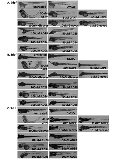
Automatically acquired images of embryos treated with increasing concentration of GSI or fragments of Gleevec. Images of embryos at 2 dpf (A), 3 dpf (B), and 5 dpf (C) were presented. Untreated embryos and 0.1% DMSO-treated embryos displayed similar phenotypes. Embryos treated with 50 μM DAPT showed a strong phenotype, and much less effect was observed at lower concentrations (5 μM and 0.5 μM). Gleevec was assessed at 100 μM, 10 μM, 1 μM, and embryos showed no phenotypic alteration. Embryos treated with 100 μM of AD95 showed morphologic changes. These changes were not obvious when 50 μM AD95 was used. |
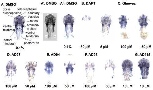
Effect of GSI and fragments of Gleevec on Notch target gene her6 expression. Embryos were treated with the compounds at 24 hpf and fixed at 48 hpf in 4% formaldehyde. In situ hybridization was performed to stain her6 staining. In control embryos, her6 expression was abundant in the dorsal diencephalon, retinas, ventral midbrain and ventral hindbrain, and it was lower in telencephalon, olfactory vesicles, branchial arches, ventral hindbrain and pectoral fins A, these regions are indicated by arrows; A′, the her6-positive brain regions from the dorsal view images were manually segmented; A″, the same embryo was illustrated for the comparison of staining intensity. B-G, her6 expression in embryos treated with different concentrations of DAPT, Gleevec, or fragments of Gleevec. |