
Image
|
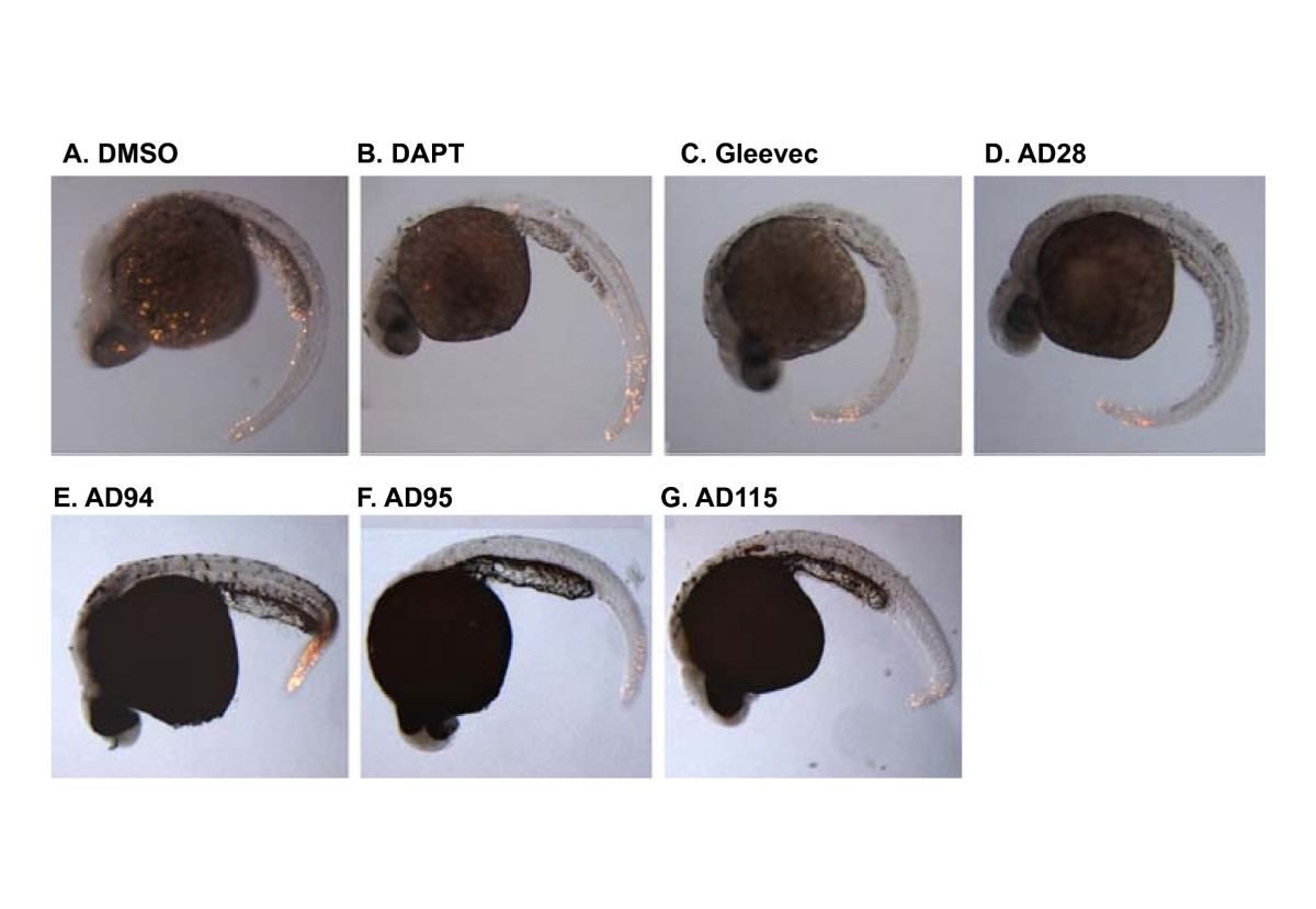
Figure Caption
Fig. 2 Apoptosis in zebrafish embryos treated with compounds. Zebrafish embryos were treated with GSI or fragments of Gleevec at 6 hpf. Embryos were then fixed and subjected to TUNEL staining at 24 hpf. Control embryos were treated with 0.1% DMSO and the remaining embryos were treated with different GSIs or Gleevec fragments at 50 μM, except for AD115 whose concentration was 10 μM. The majority of apoptotic cells were observed in the fish tail, but no difference among individual compound-treated embryos was observed. The representative images from 2 different experiments are presented.
Acknowledgments
This image is the copyrighted work of the attributed author or publisher, and
ZFIN has permission only to display this image to its users.
Additional permissions should be obtained from the applicable author or publisher of the image.
Full text @ BMC Biotechnol.