
Image
|
Figure Caption
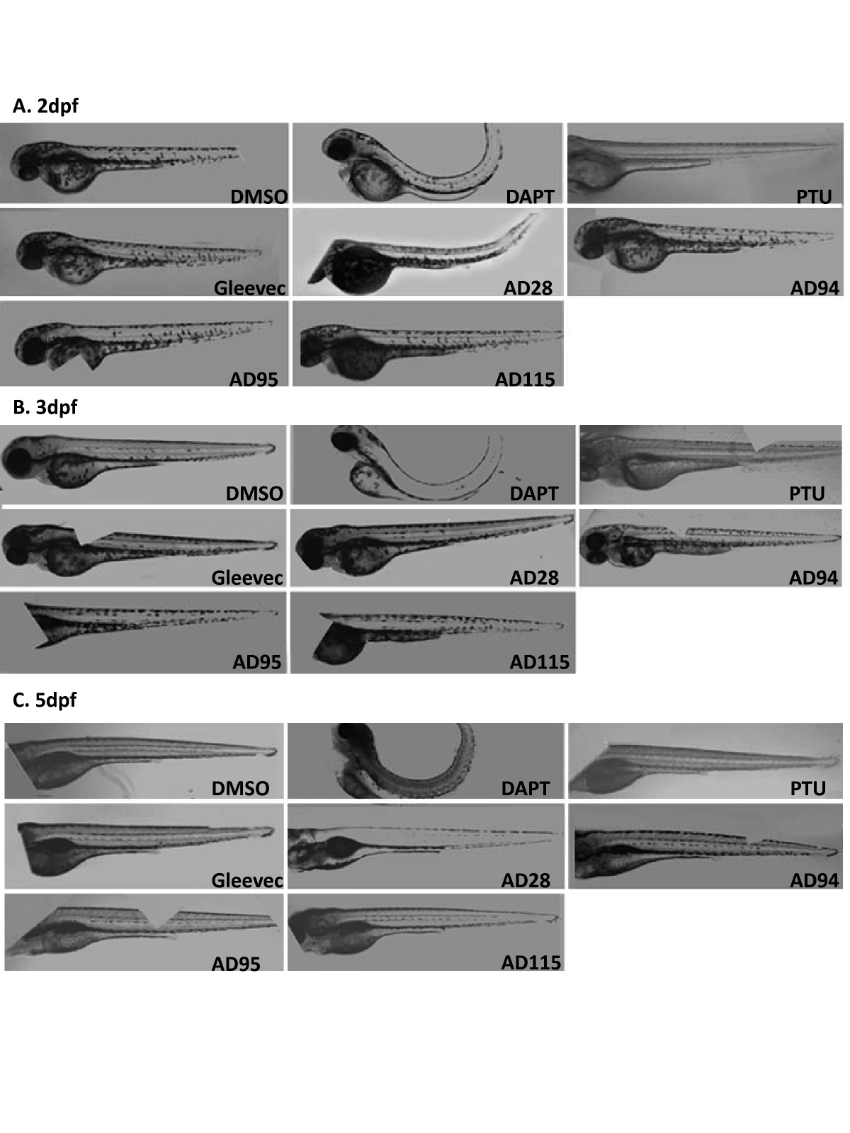
Fig. 5 Automatically acquired images of embryos treated with GSI or fragments of Gleevec at 24 hpf. Embryos were treated with GSIs and fragments of Gleevec at 24 hpf, and 50 μM of all compounds, except for 10 μM of AD115, were applied to these embryos. Images of living zebrafish in microtiter plates were acquired by In Cell Analyzer at 2 dpf (A), 3 dpf (B) and 5 dpf (C), and the same procedures were followed to automatically merge several fields of images to compose the entire zebrafish.
Acknowledgments
This image is the copyrighted work of the attributed author or publisher, and
ZFIN has permission only to display this image to its users.
Additional permissions should be obtained from the applicable author or publisher of the image.
Full text @ BMC Biotechnol.