
|
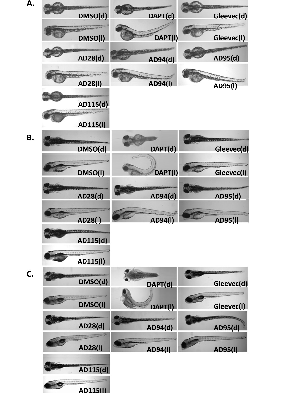
Fig. 3 Phenotypes of the embryos treated with GSI, Gleevec, and fragments at 24 hpf. Dorsal view (indicated as "d", top panel) and lateral view (indicated as "l", bottom panel) of embryos are shown. Control embryos treated with 0.1% DMSO show a wild-type phenotype. Embryos treated with GSI, DAPT, resulted in defects in somitogenesis, eye development, pigmentation, and trunk formation as observed at 2 dpf (A), 4 dpf (B), and 6 dpf (C). Embryos treated with DAPT, AD94 or AD95 started to show defects at 2 dpf, and phenotypes of remaining embryos were most obvious at 4 or 6 dpf. The following concentrations were used: 50 μM DAPT, 50 μM Gleevec, 50 μM AD28, 50 μM AD94, 50 μM AD95, and 10 μM AD115.