
- Title
-
Selective Inhibition of Retinal Angiogenesis by Targeting PI3 Kinase
- Authors
- Alvarez, Y., Astudillo, O., Jensen, L., Reynolds, A.L., Waghorne, N., Brazil, D.P., Cao, Y., O'Connor, J.J., and Kennedy, B.N.
- Source
- Full text @ PLoS One
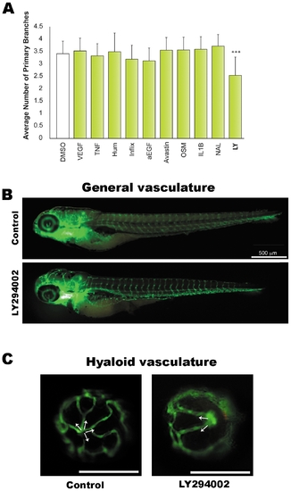
Screens reveal LY294002 to inhibit developmental angiogenesis in the eye. A. Known regulators of vasculature formation were screened for effects on developmental angiogenesis of the hyaloid vasculature in vivo. 1 day post fertilisation (dpf) Tg(fli1:EGFP) larvae were treated with the indicated drugs and the number of primary branches of hyaloid vessels determined at 5 dpf. The drugs used are: recombinant VEGF (VEGF), 50 ng/ml; Tumor Necrosis Factor (TNF), 20 ng/ml; Adalimumab - Humira® (Hum), 5 μg/ml; Infliximab (Inflix), 5 μg/ml; anti-EGF (α-EGF), 5 μg/ml; Bevacizumab- Avastin®, 5 μg/ml; Oncostatin M (OSM), 10 ng/ml; Interleukin 1 beta (IL-1B), 10 ng/ml; Nacyselyn (NAL), 10 μM; and LY294002 (LY), 10 μM. The data is plotted as the average and standard deviation of 3 replicate experiments. LY294002 results in a statistically significant inhibition of hyaloid vasculature development (***p<0.001, t-test). B–C. Fluorescent images illustrating the integrity of GFP-positive blood vessels at 5 dpf in LY294002-treated and DMSO-treated controls. No differences are observed in the morphology of the trunk vessels (B), whereas the hyaloid vessel branching pattern is abnormal and the branch number is reduced (C). White arrows in C indicate the primary branches radiating from the optic disc at the back of the lens. PHENOTYPE:
|
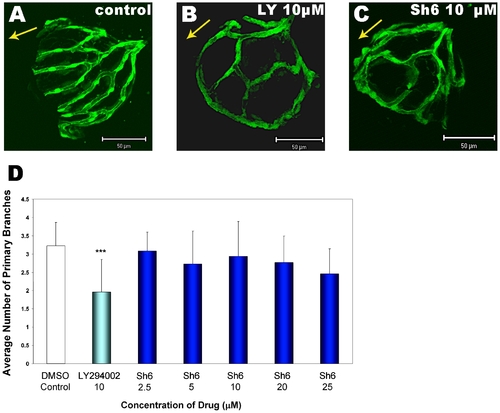
The Akt inhibitor SH6 phenocopies the PI3K inhibitor LY294002. Comparison of hyaloid vasculature patterning in DMSO-treated control larvae (A), 10 μM LY294002 treated larvae (B) and larvae treated with the Akt inhibitor SH6 at 10 μM (C). All larvae are from the Tg(fli1:EGFP) line and were treated from 1–5 dpf. Like LY294002, SH6 affects angiogenesis of the hyaloid vessels. Abnormal patterning of the hyaloid vasculature is observed with SH6 (C) resembling LY294002-treated larvae (B), though no significant reduction in primary vessel number is observed with SH6 (D). The data shown in panel D is plotted as the average and standard deviation of 3 replicate experiments (***p<0.001, t-test). PHENOTYPE:
|
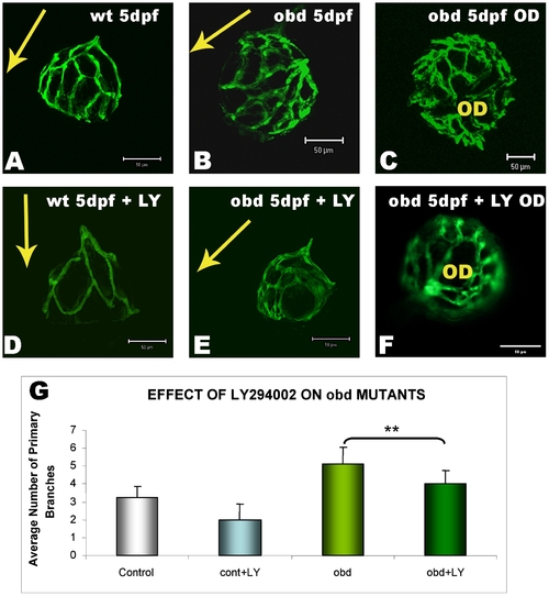
LY294002 attenuates ectopic developmental angiogenesis in obd mutants. Wildtype Tg(fli1:EGFP) larvae (wt) or out of bounds mutants in the Tg(fli1:EGFP) background (obd) were treated with 10 μM LY294002 from 1–5 dpf before dissecting lenses and analysing hyaloid vasculature development. LY294002 inhibits normal developmental angiogenesis in wildtype Tg(fli1:EGFP) larvae as seen by the reduced vessel patterning in lateral views of the lens (A, D). LY294002 also inhibits the extraneous hyaloid branch patterning (B) and branch number (C) observed in obd mutants as observed by reduction of branch patterning to near normal levels (E and F). Arrows depict the orientation of the lens, pointing in the direction from the optic disk to lens. G) Quantification of the reduction in the extraneous primary hyaloid branches number in LY294002-treated obd mutants. The data is plotted as the average and standard deviation of larvae (obd-/- n = 7, obd-/- treated: n = 8) from 2 independent experiments (***p<0.001, t-test). OD: Optic Disc. PHENOTYPE:
|
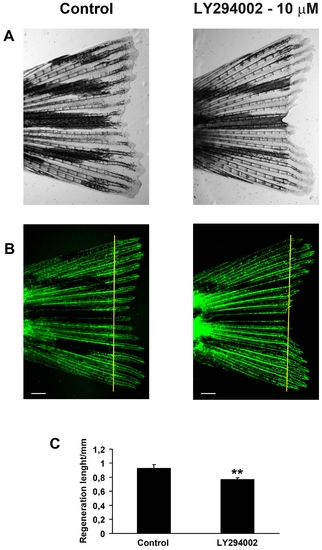
LY294002 attenuates Regenerative Angiogenesis in Adult Zebrafish. The caudal fins of adult Tg(fli1:EGFP) zebrafish were amputated and left to regenerate in either tank water or tank water supplemented with 10 μM LY294002. Bight field micrographs (A) or fluorescent micrographs (B) of representative fins at 9 days post-amputation are shown. The maximal, average blood vessel length in the regenerated tissue was quantified at the third fin ray in either fin lobe and the results are depicted in (C). LY294002 significantly inhibits regenerative angiogenesis in the tail fin (** p<0.01, t-test). Yellow lines in B indicate the amputation plane. n = 8 fin rays in 4 fish. Scale bars = 500 μm. PHENOTYPE:
|
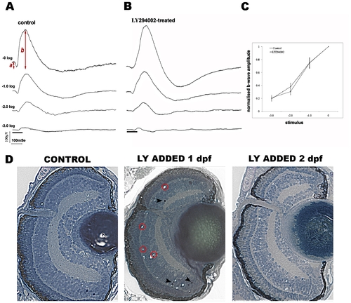
LY294002 inhibits retinal angiogenesis without affecting retinal function. A–C) Electroretinography of control (n = 13) and LY294002-treated (n = 15) zebrafish at 5 dpf. Shown is representative data from control (A) and LY294002-treated (B) larval zebrafish in response to an intensity series of white light stimulation. The stimulus flash was attenuated from 3 log units to unattenuated. The black bars at the bottom represent stimulus duration. The two groups show similar intensity response curves for the b-wave (C). To generate the intensity response curve, the average response at the maximum flash intensity was set to 1 and the average response at attenuated flash intensities was normalised to this (average ± SEM). D) Histological sections of zebrafish eyes from 5 dpf larvae treated with LY294002 from 1–5 dpf or 2–5 dpf. Retinal histology appears normal in fish treated from 2–5 dpf showing lamination, presence of optic nerve and similar size/shape to controls. In larvae treated from 1–5 dpf sparse apoptotic nuclei (arrowheads) and tissue vacuoles are observed (Magnification 400x). |
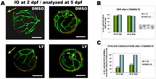
Intraocular injection of LY294002 inhibits angiogenesis but not visual function. A) The hyaloid vasculature of 5 dpf larvae intraocularly injected with 10 μM LY294002 at 2 dpf shows abnormal patterning and reduced numbers of primary branches compared with controls injected with 1% DMSO (left images are lateral views, and right images are dorsal views of lenses. OD: optic disc; scale bars: 50 μm) B) Graph of the percentage of 5 dpf larvae with optokinetic responses (OKR) following intraocular injection of LY294002 at 1 or 2 dpf. The Fisher test shows no statistically significant difference in visual function when LY294002 is injected intraocularly at 1 or 2 dpf (p values shown on table) C) Graph showing percentage of 5 dpf larvae with normal hyaloid vasculature after injection of LY294002 at 1 or 2 dpf. Intraocular LY294002 at 1 or 2 dpf, significantly inhibits hyaloid angiogenesis. Data graphed from 3 independent experiments; LY294002 at 1 dpf, n = 17; 1% DMSO at 1 dpf, n = 9; LY294002 at 2 dpf, n = 26; 1% DMSO at 2 dpf, n = 13 (***p<0.001, Chi-Square test). PHENOTYPE:
|
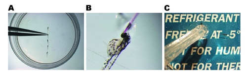
Intraocular Injection of LY294002 in zebrafish larvae. A) ∼5 dechorionated zebrafish larvae (48 hpf in the figure) are immobilized in CyGEL at RT and oriented with left eye facing upwards. Dechorionation is extremely important as non-dechorionated larvae collapse in contact with CyGEL. B) Intraocular injection (0.1–0.2 μl/eye) is performed with a heat pulled glass capillary. C) After intraocular injection the Petri dish is placed on a chilled ice pad to melt CyGEL and larvae are transferred gently but immediately to a plate with RT embryo medium for recovery. |
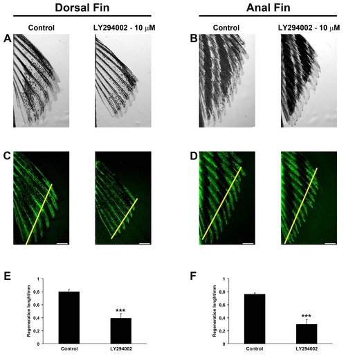
Regenerative angiogenesis in zebrafish dorsal or anal fins is inhibited by LY294002. The dorsal (A, C and E) or anal fins (B, D and F) of adult Tg(fli1:EGFP) zebrafish were amputated and left to regenerate in either tank water or tank water supplemented with 10 μM LY294002. Bight field micrographs (A and B) or fluorescent micrographs (C and D) of representative fins at 9 days post amputation are shown. The maximal, average blood vessel length in the regenerated tissue was quantified at the third fin ray from either side of each image and the results are depicted in (E and F). LY294002 significantly inhibits regenerative angiogenesis in both dorsal and anal fins (***p<0.001; t-test). Yellow lines in C and D indicate the amputation planes. n = 8 fin rays in 4 fish. Scale bars = 500 μm. |