
- Title
-
Increased DJ-1 Expression under Oxidative Stress and in Alzheimer's Disease Brains
- Authors
- Baulac, S., Lu, H., Strahle, J., Yang, T., Goldberg, M.S., Shen, J., Schlossmacher, M.G., Lemere, C.A., Lu, Q., and Xia, W.
- Source
- Full text @ Mol. Neurodegener.
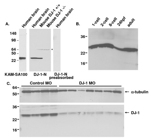
Knockdown of DJ-1 in zebrafish embryos. A. Abundant DJ-1 expression in human brain lysate from a control case, as detected by Western Blot (WB) with KAM-SA100 and anti-DJ-1-N antibodies, but not when anti-DJ-1-N was pre-absorbed with the synthetic DJ-1 peptide. Specificity of anti-DJ-1-N antibody was confirmed by detecting DJ-1 in wild type mouse brain lysate but not in the DJ-1 knockout mouse brain lysate. A minor cross-reacting band at ∼70 kDa was detected in extracts of both wild type and DJ-1 knockout mouse brain, but not in human brain extracts. B. Zebrafish embryos at different developmental stages and brains of adult zebrafish were lysed for Western blotting with antibody E2.19. High levels of DJ-1 protein were found in embryos at all developmental stages and in adult brains. C. Extracts taken from individual embryos at 24 hpf were run on WB with antibody E2.19 (bottom panel). The same blot was re-probed with antibody against α-tubulin (top panel). DJ-1 protein levels were dramatically reduced in DJ-1 KD embryos compared to control embryos. |
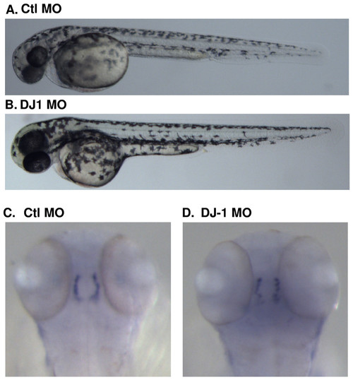
DJ-1 expression is not essential for proper zebrafish development. A, B. Embryos were injected with control or DJ-1 MO at the one cell stage, and images were acquired at 48 hpf. DJ-1 KD fish did not differ in morphologic phenotype compared to control MO injected zebrafish. C, D. Embryos injected with control or DJ-1 MO were fixed at 48 hpf, followed by in situ hybridization using a probe against TH. TH staining of control or DJ-1 MO injected embryos was almost identical. |
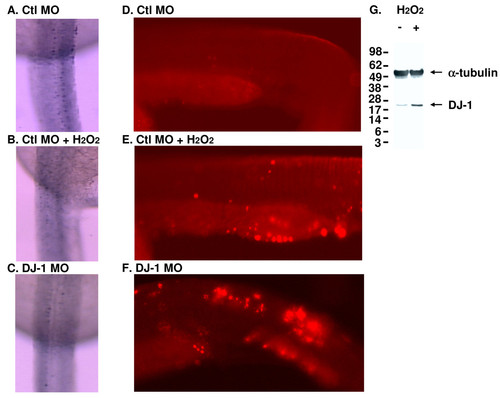
DJ-1 expression is induced in zebrafish treated with H2O2 and knockdown of DJ-1 increases the number of apoptotic cells. A. Control MO injected embryos showed normal distribution of islet-1 positive neurons along the spinal cord, as illustrated by the dorsal view of the spinal cord, anterior to the top of the image. B. Control MO injected embryos treated with H2O2 showed a slight reduction in islet-1 staining. C. DJ-1 MO injected embryos displayed even weaker islet-1 staining. D-F. Control MO (D, E) or DJ-1 MO (F) was injected into embryos at one cell stage; after 24 hr, control MO injected embryos were treated with 0.03% H2O2 for 30 min. Compared to control MO injected embryos (D), there was an increase of TUNEL positive cells in the tails of H2O2 treated embryos (E) and DJ-1 MO injected embryos (F). The lateral view of the trunk region of zebrafish was illustrated with anterior to the left of the image. The number of apoptotic cells in the tails of H2O2 treated embryos (E) and DJ-1 KD embryos (F) is higher than that in control MO injected embryos (D). G. Adult zebrafish were treated with 0.03% H2O2 for 30 min before brains were harvested and lysed for Western blot. α-Tubulin (an internal sample loading control) was detected over the upper portion of the blot, and the bottom portion of the blot was detected with antibody DJ-1-N. While the levels of α-tubulin were not changed in the presence of H2O2, the levels of DJ-1 were increased. |