
- Title
-
Expression of multiple class three semaphorins in the retina and along the path of zebrafish retinal axons
- Authors
- Callander, D.C., Lamont, R.E., Childs, S.J., and McFarlane, S.
- Source
- Full text @ Dev. Dyn.
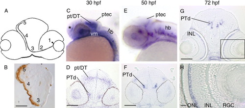
Expression of sema3Aa. A: Schematic diagram of a transverse section of the optic pathway. A RGC resides in the RGC layer (1) and sends out an axon that exits the retina at the optic nerve head (2), crosses the midline at the optic chiasm (3), extends along the pial surface of the diencephalon (4), and enters the main target, the optic tectum (5). B: Transverse section of a 72-hpf brain with an HRP-labelled RGC optic projection (brown). The eyes and skin have been removed. The optic chiasm, pathway, and tectum are labelled 3, 4, and 5, respectively. C and E are lateral views of whole-mount embryos and D and F are stage-matched transverse sections. C, D: sema3Aa transcripts appear in the pretectum/dorsal thalamus and the posterior tectum at 30 hpf. Bilateral nuclei in the dorsal posterior tuberculum also show expression. From 30-72 hpf, sema3Aa transcripts are seen in the telencephalon (asterisk), the hindbrain, and the ventral mesenchymal tissue. E, F: At 50 hpf, signal is maintained in the dorsal posterior tuberculum (F) and the posterior tectum (E). G, H: At 72 hpf, in transverse sections the outer region of the inner nuclear layer begins to express sema3Aa. The boxed area in G is shown at a higher magnification in H. hb, hindbrain; INL, inner nuclear layer; ONL, outer nuclear layer; ptec, posterior tectum; pt/DT, pretectum/dorsal thalamus; PTd, dorsal posterior tuberculum; RGC, retinal ganglion cell layer; vm, ventral mesenchyme. Scale bars = 50μm (B,D,F,G), 10 μm (H). EXPRESSION / LABELING:
|
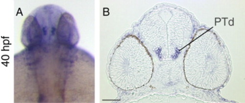
Expression of sema3C. A, B: A dorsal view of a whole-mount embryo is shown in A, and a transverse section through the eyes and brain is shown in B. sema3C transcripts are seen in the dorsal posterior tuberculum at 40 hpf. PTd, dorsal posterior tuberculum. Scale bar in B = 50 μm. |
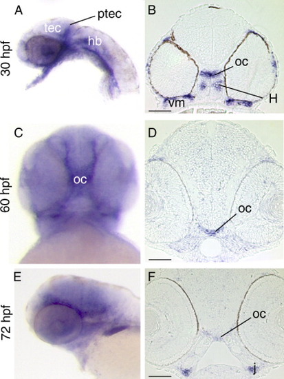
Expression of sema3E. Lateral (A,E) and ventral views (C) of whole-mount embryos, and stage-matched transverse sections (B,D,F). A: sema3E label is seen in the posterior tectum at 30 hpf. B,D,F: sema3E transcripts appear in the optic chiasm at 30 hpf and persist until 72 hpf. sema3E is seen in the ventral mesenchymal tissue (B), the hypothalamus (B), and the jaw (F). tec, tectum; hb, hindbrain; H, hypothalamus; j, jaw; oc, optic chiasm; ptec, posterior tectum; vm, ventral mesenchyme. Scale bars in B,D,F = 50 μm. |
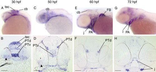
Expression of sema3Fa. A,C,E, and G are lateral views of whole-mount embryos and B,D,F, and H are stage-matched transverse sections. A,B: At 30 hpf, sema3Fa transcript is seen in the ventral retina (arrowhead), in the optic tectum, and in the ventral and dorsal regions of the posterior tuberculum. There is also label in the cerebellum. C,D: At 50 hpf, sema3Fa is expressed in the ventral posterior tuberculum and in bilateral nuclei in the dorsal posterior tuberculum. Expression in the ventral retina (arrowhead) continues. E,F: At 60 hpf, expression in the dorsal posterior tuberculum and cerebellum is maintained. G,H: At 72 hpf, there is diffuse label in the anterior brain (H). There is also expression in the jaw (E,G,H), pharyngeal arches (E,G). and the pectoral fin buds (E). cb, cerebellum; FB, fin bud; j, jaw; PA, pharyngeal aches; PTd, dorsal posterior tuberculum; PTv, ventral posterior tuberculum; tec, tectum. Scale bars in B,D,F, H = 50 μm. EXPRESSION / LABELING:
|
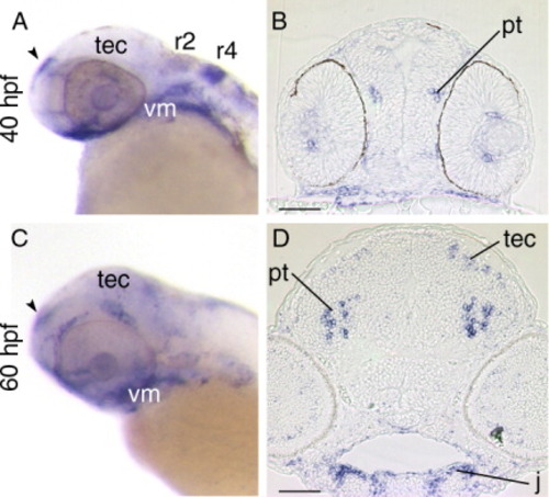
Expression of sema3Fb. A,C: Lateral views of whole-mount embryos. B,D: Stage-matched transverse sections. B,D: At 40-60 hpf, the pretectal region of the diencephalon expresses sema3Fb transcripts. C,D: At 60 hpf, the region of the tectum just ventral to where the RGC axons terminate expresses sema3Fb. The jaw (D), ventral mesenchyme (A,C), telencephalon (arrowhead; A,C), and rhombomeres 2 and 4 (A) also show expression. j, jaw; pt, pretectum; r, rhombomere; tec, tectum; vm, ventral mesenchyme. Scale bars in B,D = 50 μm. |
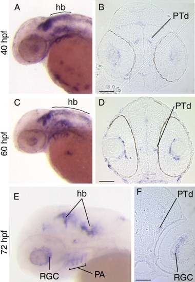
Expression of sema3Ga. A,C,E: Lateral views of whole-mount embryos. B,D,F: Stage-matched transverse sections. A,B: At 40 hpf, sema3Ga expression is seen in the dorsal posterior tuberculum as well as in the dorsal hindbrain. Blood vessels bordering the lens are also stained at 40 (B) and 60 (D) hpf. C,D: The dorsal posterior tuberculum expression persists at 60 hpf. E,F: Expression is seen in the RGC layer at 72 hpf (E, F), whereas signal in the dorsal hindbrain has switched to two ventrolateral regions (compare C and E). sema3Ga transcripts are present in the dorsal posterior tuberculum from 40-60 hpf (B,D,F) and in the pharyngeal arches at 72 hpf (E). hb, hindbrain; PA, pharyngeal arches; PTd, dorsal posterior tuberculum; RGC, retinal ganglion cell layer. Scale bars in B,D,F = 50 μm. EXPRESSION / LABELING:
|
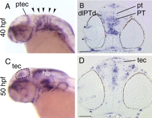
Expression of sema3Gb. A,C: Lateral views of whole-mount embryos. B,D: Stage-matched transverse sections. A: At 40 hpf, in situ signal is evident in the posterior tectum and the rhombomeric boundaries (arrowheads). B: At 40 hpf, sema3Gb is expressed in the peripheral retina (asterisk). Several areas of the diencephalon express sema3Gb: dorsolateral region of the dorsal posterior tuberculum, the dorsoventral axis of the posterior tuberculum, and the pretectal area. C,D: At 50 hpf, a subset of cells dispersed throughout the tectum express sema3Gb, which is evident in the whole-mount embryo (dashed line outlines the tectum) and in the section. pt, pretectum; ptec, posterior tectum; tec, tectum; PT, posterior tuberculum; dlPTd, dorsolateral region of the dorsal posterior tuberculum; Scale bars in B,D = 50 μm. |