
Image
|
Figure Caption
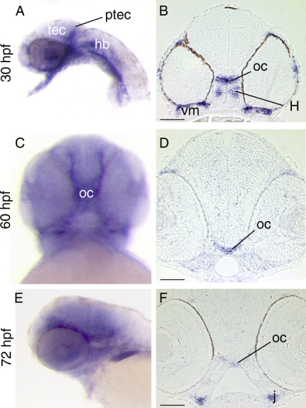
Fig. 3 Expression of sema3E. Lateral (A,E) and ventral views (C) of whole-mount embryos, and stage-matched transverse sections (B,D,F). A: sema3E label is seen in the posterior tectum at 30 hpf. B,D,F: sema3E transcripts appear in the optic chiasm at 30 hpf and persist until 72 hpf. sema3E is seen in the ventral mesenchymal tissue (B), the hypothalamus (B), and the jaw (F). tec, tectum; hb, hindbrain; H, hypothalamus; j, jaw; oc, optic chiasm; ptec, posterior tectum; vm, ventral mesenchyme. Scale bars in B,D,F = 50 μm.
Figure Data
Acknowledgments
This image is the copyrighted work of the attributed author or publisher, and
ZFIN has permission only to display this image to its users.
Additional permissions should be obtained from the applicable author or publisher of the image.
Full text @ Dev. Dyn.