
|
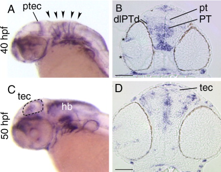
Fig. 7 Expression of sema3Gb. A,C: Lateral views of whole-mount embryos. B,D: Stage-matched transverse sections. A: At 40 hpf, in situ signal is evident in the posterior tectum and the rhombomeric boundaries (arrowheads). B: At 40 hpf, sema3Gb is expressed in the peripheral retina (asterisk). Several areas of the diencephalon express sema3Gb: dorsolateral region of the dorsal posterior tuberculum, the dorsoventral axis of the posterior tuberculum, and the pretectal area. C,D: At 50 hpf, a subset of cells dispersed throughout the tectum express sema3Gb, which is evident in the whole-mount embryo (dashed line outlines the tectum) and in the section. pt, pretectum; ptec, posterior tectum; tec, tectum; PT, posterior tuberculum; dlPTd, dorsolateral region of the dorsal posterior tuberculum; Scale bars in B,D = 50 μm.