
- Title
-
Analysis of zebrafish sidetracked mutants reveals a novel role for Plexin A3 in intraspinal motor axon guidance
- Authors
- Palaisa, K.A., and Granato, M.
- Source
- Full text @ Development
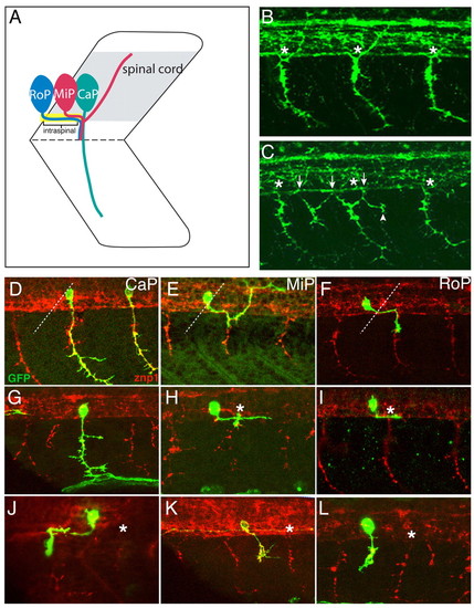
Motoneuronal defects in sidetracked mutants. (A) Schematic of primary motoneurons. (B) 24 hpf wild-type embryos stained with SV2. Endogenous exit points are labeled with asterisks. (C) 24 hpf sidetracked (set) embryos stained with SV2. Endogenous exit points are labeled with asterisks, ectopic exit points are marked by arrows, and branches by an arrowhead. (D-F) Wild-type embryos injected with Hb9:GFP plasmid and stained with anti-GFP (green) and SV2 (red) to visualize CaP, MiP and RoP trajectories. Broken lines indicate the position of somite boundaries. (G-L) set embryos injected with Hb9:GFP plasmid and processed as above. Notice that the labeled somata are located at a distance from the endogenous exit point (asterisk). (G) Normal projecting CaP. (H,I) Example of MiP/RoP neurons bypassing the exit point. (J) Rostrally projecting MiP/RoP motoneurons. Notice that growth cones exit the spinal cord through the adjacent, rostral exit point. (K,L) Examples of ectopic-exiting RoP/MiP axons. CaP, caudal primary; MiP, middle primary; RoP, rostral primary. PHENOTYPE:
|
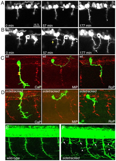
Time-lapse analysis and branching. (A) Individual movie frames (see Movie 1 in the supplementary material) showing wild-type CaP/VaP migration. Notice the growth cone from an interneuron extending along the ventral aspect of the spinal cord (arrowhead). (B) Individual movie frames (see Movie 2 in the supplementary material) showing the ectopic exit of a sidetracked MiP/RoP axon (asterisk). The soma is located between two CaP/VaP pairs. Mutant growth cones pioneer an ectopic exit zone into the periphery, where they often branch (yellow arrow) and sometimes join endogenous motor axons (white arrow). Notice the growth cone from an interneuron extending along the ventral aspect of the spinal cord (arrowhead). sidetracked mutants display excessive branching. (C,D) 24 hpf embryos injected with Hb9:GFP plasmid, and stained with anti-GFP (green) and SV2 (red) to visualize CaP, MiP and RoP trajectories, respectively. (C) Wild-type (wt) embryo. (D) sidetracked embryo. Notice the small branches along the axonal shaft. (E,F) 48 hpf wild-type and sidetracked embryos stained with znp1 and SV2. Notice the persisting ectopic exit points (arrows) and the long branches (arrowheads) present in the mutants. CaP, caudal primary; MiP, middle primary; RoP, rostral primary. PHENOTYPE:
|
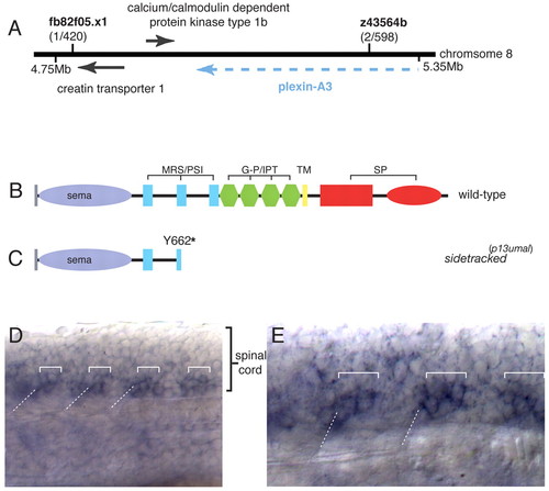
Molecular cloning and plexin A3 expression. (A) Molecular genetic map of the sidetracked (plexin A3) region. (B) Domain organization of the wild-type Plexin A3 protein. (C) The sidetrackedp13umal allele is a presumptive null. For domains, see main text. (D) Lateral view of a wild-type embryo processed for plexin A3 in situ hybridization. Low-level expression is detectable throughout the spinal cord and appears enriched in neurons close to the somite boundary. (E) High-magnification view. Expression of plexin A3 is enriched in two somata adjacent to the somite boundary, consistent with the positions of middle primary (MiP) and rostral primary (RoP). Square brackets indicate motoneuron cell bodies; broken lines indicate the position of somite boundaries. G-P/IPT, glycine-proline-rich/immunoglobulin-like fold shared by plexins; MRS/PSI, Met-related sequence/plexin-semaphorin-integrin domain; SP, Sex-Plexin; TM, transmembrane. EXPRESSION / LABELING:
|
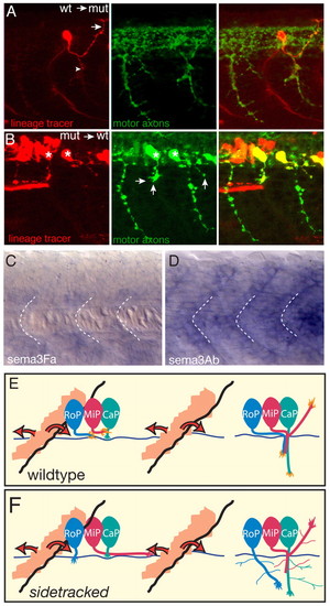
Plexin A3 acts cell autonomously, mediating the chemorepulsion of motor axon growth cones from posterior somites by semaphorin ligands. (A,B) Chimera analysis. Donor cells are labeled with rhodamine dextran (red) and donor motoneurons express GFP (green); hosts were also stained with SV2 antibody (green) to reveal all motor axonal trajectories. (A) A wild-type (wt) donor-derived MiP neuron (red) in an otherwise mutant (mut) host properly exits the spinal cord, projects to the choice point (arrowhead), and then forms a dorsal collateral. Arrow indicates normal dorsal trajectory. (B) sidetracked donor-derived motoneurons (red, asterisks) in an otherwise wild-type host exit the spinal cord ectopically (arrows). (C) sema3Fa expression at 21 hpf. Broken lines indicate the position of somite boundaries. (D) sema3Ab expression at 21 hpf. (E) Model of wild-type Plexin A3 function. During intraspinal motor axon guidance, a diffusible repulsive cue (semaphorin; orange), secreted from the posterior somite, spreads anteriorly and posteriorly. This repulsion directs Plexin A3-sensitive growth cones (RoP, MiP) towards the mid-segmental exit zone. After they exit from the spinal cord, CaP, MiP and RoP motoneurons navigate towards their respective muscle targets. (F) In sidetracked mutants, MiP and RoP motoneurons are insensitive to semaphorin repulsion during intraspinal guidance and extend aberrantly. After they exit from the spinal cord and navigate towards their targets, mutant CaP, MiP and RoP motoneurons exhibit exuberant branching, suggesting that Plexin A3 signaling is also important to prevent precocious spreading of motor axonal branches. CaP, caudal primary; MiP, middle primary; RoP, rostral primary. EXPRESSION / LABELING:
|