
|
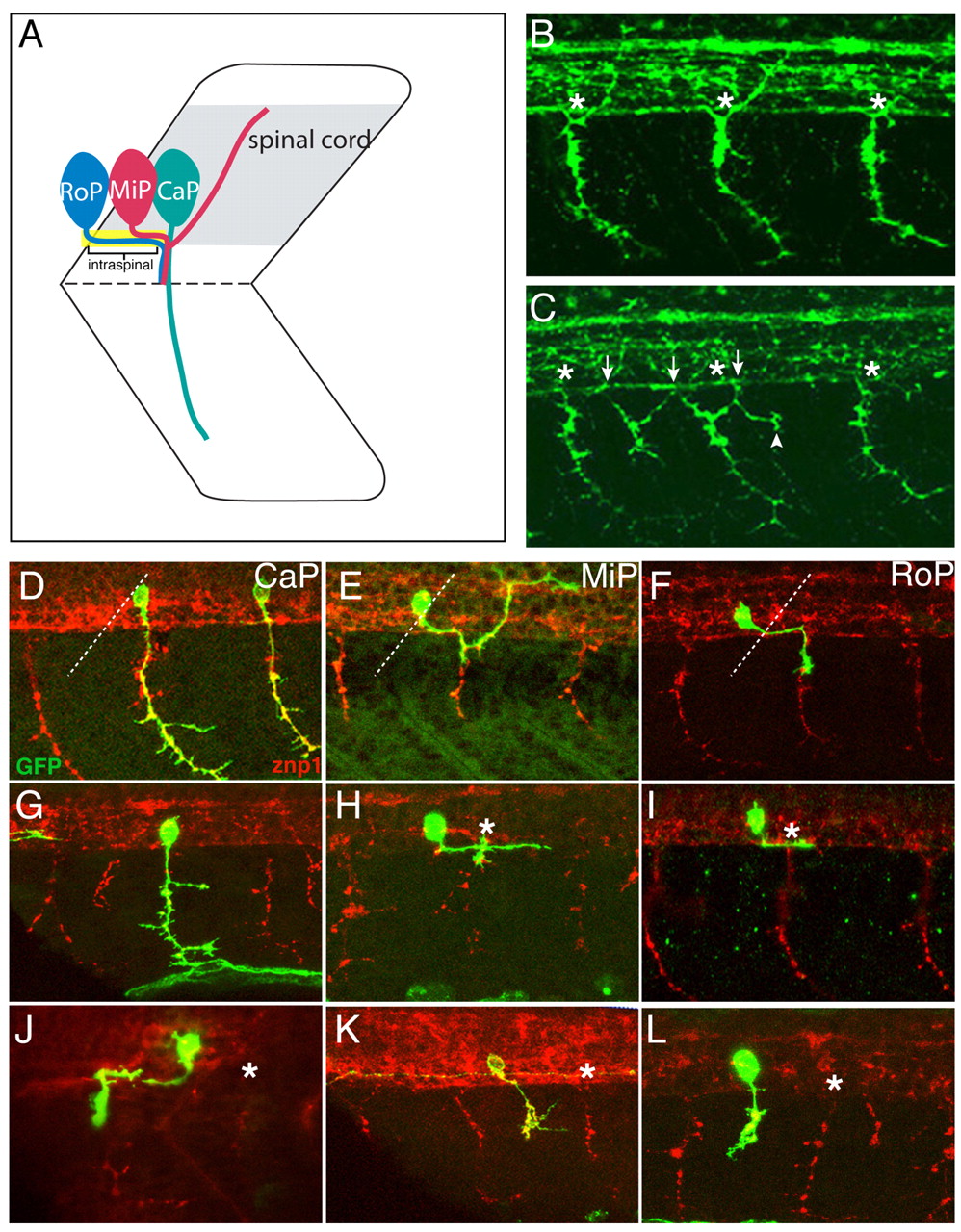
Fig. 1 Motoneuronal defects in sidetracked mutants. (A) Schematic of primary motoneurons. (B) 24 hpf wild-type embryos stained with SV2. Endogenous exit points are labeled with asterisks. (C) 24 hpf sidetracked (set) embryos stained with SV2. Endogenous exit points are labeled with asterisks, ectopic exit points are marked by arrows, and branches by an arrowhead. (D-F) Wild-type embryos injected with Hb9:GFP plasmid and stained with anti-GFP (green) and SV2 (red) to visualize CaP, MiP and RoP trajectories. Broken lines indicate the position of somite boundaries. (G-L) set embryos injected with Hb9:GFP plasmid and processed as above. Notice that the labeled somata are located at a distance from the endogenous exit point (asterisk). (G) Normal projecting CaP. (H,I) Example of MiP/RoP neurons bypassing the exit point. (J) Rostrally projecting MiP/RoP motoneurons. Notice that growth cones exit the spinal cord through the adjacent, rostral exit point. (K,L) Examples of ectopic-exiting RoP/MiP axons. CaP, caudal primary; MiP, middle primary; RoP, rostral primary.