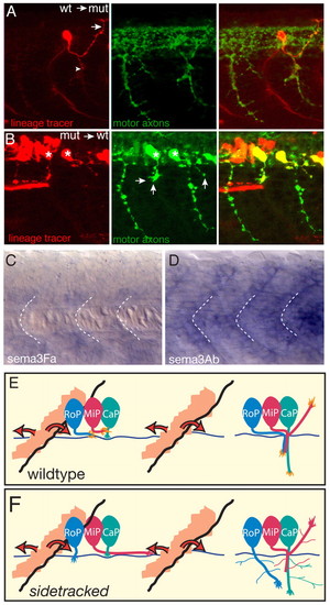
Plexin A3 acts cell autonomously, mediating the chemorepulsion of motor axon growth cones from posterior somites by semaphorin ligands. (A,B) Chimera analysis. Donor cells are labeled with rhodamine dextran (red) and donor motoneurons express GFP (green); hosts were also stained with SV2 antibody (green) to reveal all motor axonal trajectories. (A) A wild-type (wt) donor-derived MiP neuron (red) in an otherwise mutant (mut) host properly exits the spinal cord, projects to the choice point (arrowhead), and then forms a dorsal collateral. Arrow indicates normal dorsal trajectory. (B) sidetracked donor-derived motoneurons (red, asterisks) in an otherwise wild-type host exit the spinal cord ectopically (arrows). (C) sema3Fa expression at 21 hpf. Broken lines indicate the position of somite boundaries. (D) sema3Ab expression at 21 hpf. (E) Model of wild-type Plexin A3 function. During intraspinal motor axon guidance, a diffusible repulsive cue (semaphorin; orange), secreted from the posterior somite, spreads anteriorly and posteriorly. This repulsion directs Plexin A3-sensitive growth cones (RoP, MiP) towards the mid-segmental exit zone. After they exit from the spinal cord, CaP, MiP and RoP motoneurons navigate towards their respective muscle targets. (F) In sidetracked mutants, MiP and RoP motoneurons are insensitive to semaphorin repulsion during intraspinal guidance and extend aberrantly. After they exit from the spinal cord and navigate towards their targets, mutant CaP, MiP and RoP motoneurons exhibit exuberant branching, suggesting that Plexin A3 signaling is also important to prevent precocious spreading of motor axonal branches. CaP, caudal primary; MiP, middle primary; RoP, rostral primary.
|
