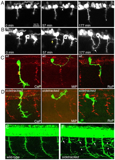
Time-lapse analysis and branching. (A) Individual movie frames (see Movie 1 in the supplementary material) showing wild-type CaP/VaP migration. Notice the growth cone from an interneuron extending along the ventral aspect of the spinal cord (arrowhead). (B) Individual movie frames (see Movie 2 in the supplementary material) showing the ectopic exit of a sidetracked MiP/RoP axon (asterisk). The soma is located between two CaP/VaP pairs. Mutant growth cones pioneer an ectopic exit zone into the periphery, where they often branch (yellow arrow) and sometimes join endogenous motor axons (white arrow). Notice the growth cone from an interneuron extending along the ventral aspect of the spinal cord (arrowhead). sidetracked mutants display excessive branching. (C,D) 24 hpf embryos injected with Hb9:GFP plasmid, and stained with anti-GFP (green) and SV2 (red) to visualize CaP, MiP and RoP trajectories, respectively. (C) Wild-type (wt) embryo. (D) sidetracked embryo. Notice the small branches along the axonal shaft. (E,F) 48 hpf wild-type and sidetracked embryos stained with znp1 and SV2. Notice the persisting ectopic exit points (arrows) and the long branches (arrowheads) present in the mutants. CaP, caudal primary; MiP, middle primary; RoP, rostral primary.
|
