
- Title
-
T-box binding sites are required for activity of a cardiac GATA-4 enhancer
- Authors
- Heicklen-Klein, A., and Evans, T.
- Source
- Full text @ Dev. Biol.
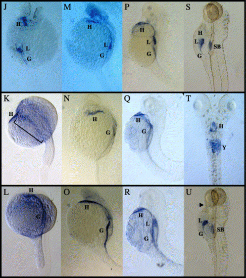
Whole-mount in situ hybridization analysis of GATA-4/5/6. (A–C) Dorsal views of five somite (12 hpf) embryos, anterior at the top, showing GATA-4 (A), GATA-5 (B) and GATA-6 (C) expression in the lateral plate mesoderm (LPM). (D–F) Dorsal views of 10 somite (14 hpf) embryos, anterior at the top, showing GATA-4 (D), GATA-5 (E) and GATA-6 (F) expression. All three GATA factors are expressed in the LPM. However, GATA-4 is also expressed in the tail bud (*) as previously reported in zebrafish (Griffin et al., 2000), GATA-5 expression is lower in the central portion of the LPM (denoted by a line) and GATA-6 is expressed posteriorly in the region medial to the stripes of the LPM (arrow) as previously shown in mouse (Morrisey et al., 1996). D is rotated slightly relative to E and F to show tail bud expression. (G–I) Dorsal views of embryos at 18 hpf, anterior at the top, showing GATA-4 (G), GATA-5 (H) and GATA-6 (I) expression in the cardiac progenitors migrating to the ventral aspect of the embryo. GATA-5 retains expression in posterior regions of the LPM (bracket) as shown previously in Xenopus (Jiang and Evans, 1996). (J–L) Dorsal views at 24 hpf, anterior at the top. GATA-4 (J), GATA-5 (K) and GATA-6 (L) expression can be seen in the fused heart tube. GATA-4 is also expressed in the liver, GATA-5 continues to have low levels of expression in the posterior LPM (bracket) and GATA-6 is expressed in a broad region of the gut tube. (M–O) Lateral views at 28 hpf, anterior at the top, showing GATA-4 (M), GATA-5 (N) and GATA-6 (O) expression at the beating heart stage. GATA-4 expression remains in the liver but now extends to more extensive regions of the gut. GATA-5 expression in the gut is just visible and restricted to the central portion. GATA-6 displays the most extensive pattern of gut expression but does not appear to be expressed at high levels in the liver at this stage. (P–R) Lateral views at 48 hpf, anterior at the top, showing GATA-4 (P), GATA-5 (Q) and GATA-6 (R) expression in the heart and gut tube. (S, U) Lateral and (T) ventral views at 6 days post fertilization, anterior at the top, showing GATA-4 (S) expression in the heart and gut, GATA-5 (T) expression in the heart, and GATA-6 (U) expression in the gut, heart and two of the posterior pharyngeal pouches (arrow). Stainings in the swim bladder in (S) and (U) and in the yolk in (T) are nonspecific staining artifacts. G = gut, H = heart, L = liver, SB = swim bladder and Y = yolk. EXPRESSION / LABELING:
|
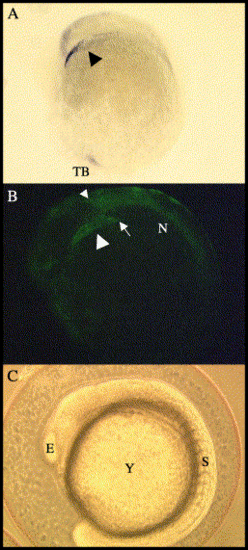
The GATA-4 promoter recapitulates the endogenous expression pattern during cardiogenesis. (A) In situ hybridization of GATA-4 RNA stains a stripe of cells just dorsal to the yolk (large black arrowhead). (B) GFP expression controlled by the 14.8-kb GATA-4 promoter in the heart also forms a similar stripe in the LPM resembling that in A (large white arrowhead). Note that the posterior extent of the GFP-positive heart precursors (arrow) overlaps with the anterior extend of GFP-positive cells in the notochord (small arrowhead). (C) Phase image corresponding to B. All images are lateral views. E = eye, N = notochord, S = somites, TB = tailbud and Y = yolk. EXPRESSION / LABELING:
|
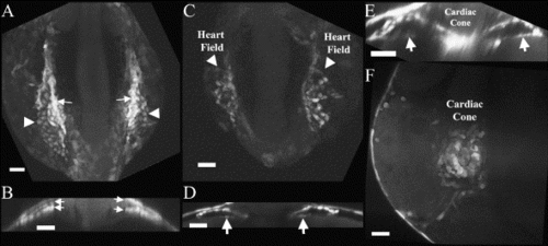
GATA-4 upstream sequence (14.8 kb) regulates the GFP expression pattern during cardiac cone fusion. (A) At 10 somites (14 hpf), the heart field is visible on the lateral aspects of the zebrafish embryo as clusters of GFP-positive cells. Note that the cells in the LPM appear to express GFP at two different intensities, a central population of cells (arrows) and the less intensely labeled lateral population of cells (arrowhead). (B) Cross section at this stage through the central region of these stripes reveals two layers of GFP-positive cells (arrows). (C) At 16 hpf, two domains of GFP-positive cells can be seen on either side of the embryo. (D) Cross section through the central portion of these heart fields shows two tubes just before migration to the ventral midline (arrows). (E) Cross section at 18 hpf at the level where the cone has begun to fuse demonstrates that the two GFP heart fields form tubes (arrows) that are fusing at the midline to become the cardiac cone. (F) By 20 hpf, transgene-regulated GFP-positive cells have fused to form the cardiac cone. A, C and F are dorsal projections created from images using confocal or multiphoton microscopes. Scale BAR = 50 μM. |
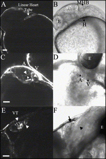
Visualization of the developing heart tube using 14.8 kb of GATA-4 regulatory sequences to direct GFP expression. (A) GFP expression is seen throughout the newly fused heart tube at 24 hpf. (B) Phase of A. (C) GFP expression is seen in the endocardium (arrowheads) and the myocardium (arrows) of both the atrium and the ventricle at 48 hpf. (D) Phase of C. (E) GFP expression is present in the ventricular trabeculae (arrow) and the bulboventricular valve (arrow head) of the heart at 6 days post fertilization. (F) Phase of E. A, B, E and F are lateral views with anterior to the left. C and D are ventral views with the head up. Scale BAR = 50 μM. A = atrium, E = eye, H = heart, MHB = midbrain hindbrain border, V = ventricle and VT = ventricular trabeculae. |
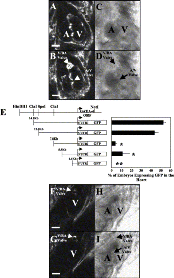
Distal elements of the GATA-4 promoter are required to direct expression to the atrioventricular valve and caudal aspects of the heart at 6 days post fertilization. (A, B) The 14.8-kb GATA-4 fragment controls GFP expression in the atrioventricular valve (A/V Valve), the bulboventricular valve (V/BA Valve), the ventricle and the atrium. A and B are images at different focal planes to enable visualization of specific regions of the three-dimensional heart. (C, D) Phase of A and B, respectively. (E) GATA-4 promoter analysis. Transient transgenic assays showed that 14.8 or 12 kb of 5′-flanking GATA-4 genomic DNA confers GFP expression to the heart. The HindIII–NotI genomic fragment used to clone the various GATA-4 constructs is schematically represented at the top of the graph. Truncation of 4.2 kb from the 12-kb construct significantly decreased GFP expression in the heart (7.8-kb construct). GFP expression in the heart is lost following further truncation to 1.1 kb of sequence. Statistical analysis was performed between 4 and 6 days post fertilization. Student's t test was performed in comparison to the 12-kb promoter fragment and represents a significant decrease (P < 0.01) in the ability of a specific construct to direct GFP expression in the heart. Significance is indicated by an *. No expression in the heart is indicated by **. Significance could not be determined for constructs that do not express GFP in the heart because the analysis requires standard deviation. However, there is a difference in the expression level between the 1.1-kb construct and the other constructs. ORF = open reading frame. (F, G) Comparable images of A and B except that the transgene contains only 7.8 kb of GATA-4 upstream sequences. Note that expression in the heart is limited to the ventricle and the bulboventricular valve (V/BA valve). F and G are images at different focal planes to enable visualization of specific regions of the three-dimensional heart. (H, I) Phase of F and G, respectively. All images are ventral views with the head at the top of the picture. Scale BAR = 50 μM. A = atrium and V = ventricle. |
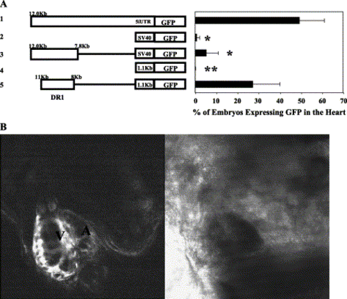
A 3-kb distal element upstream of the proximal promoter region is sufficient to drive expression in both chambers of the heart. (A) Neither the SV40 promoter (construct 2) nor the proximal promoter (construct 4) was capable of providing significant levels of GFP expression in the heart, compared to 12 kb of upstream genomic sequences (construct 1). Similarly, the 4.2-kb distal fragment placed upstream of the SV40 minimal promoter was not sufficient to confer significant GFP expression to the heart (construct 3). However, a subregion of the 4.2-kb sequence containing 3 kb of GATA-4 DNA upstream of the 1.1-kb proximal region of the GATA-4 promoter was sufficient to confer GFP expression to the heart (construct 5). Student's t test was performed in comparison to the 12-kb promoter fragment and a significant difference (P < 0.01) in the ability of a construct to drive GFP expression in the heart is indicated as in Fig. 5. (B) Confocal analysis of an embryo transgenic for the 3-kb distal region from -11 to -8 kb together with the 1.1-kb proximal region of the GATA-4 promoter (germline transmission of construct 5 in A). GFP is expressed in both chambers of the heart recapitulating the results found in A. |
Reprinted from Developmental Biology, 267(2), Heicklen-Klein, A., and Evans, T., T-box binding sites are required for activity of a cardiac GATA-4 enhancer, 490-504, Copyright (2004) with permission from Elsevier. Full text @ Dev. Biol.