
|
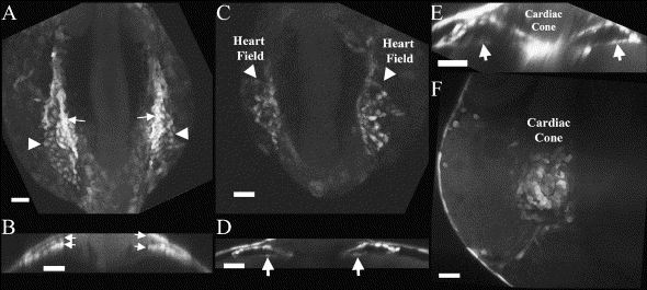
Fig. 3 GATA-4 upstream sequence (14.8 kb) regulates the GFP expression pattern during cardiac cone fusion. (A) At 10 somites (14 hpf), the heart field is visible on the lateral aspects of the zebrafish embryo as clusters of GFP-positive cells. Note that the cells in the LPM appear to express GFP at two different intensities, a central population of cells (arrows) and the less intensely labeled lateral population of cells (arrowhead). (B) Cross section at this stage through the central region of these stripes reveals two layers of GFP-positive cells (arrows). (C) At 16 hpf, two domains of GFP-positive cells can be seen on either side of the embryo. (D) Cross section through the central portion of these heart fields shows two tubes just before migration to the ventral midline (arrows). (E) Cross section at 18 hpf at the level where the cone has begun to fuse demonstrates that the two GFP heart fields form tubes (arrows) that are fusing at the midline to become the cardiac cone. (F) By 20 hpf, transgene-regulated GFP-positive cells have fused to form the cardiac cone. A, C and F are dorsal projections created from images using confocal or multiphoton microscopes. Scale BAR = 50 μM.
Reprinted from Developmental Biology, 267(2), Heicklen-Klein, A., and Evans, T., T-box binding sites are required for activity of a cardiac GATA-4 enhancer, 490-504, Copyright (2004) with permission from Elsevier. Full text @ Dev. Biol.