
Fig. 1
- ID
- ZDB-FIG-051122-2
- Publication
- Heicklen-Klein et al., 2004 - T-box binding sites are required for activity of a cardiac GATA-4 enhancer
- Other Figures
- All Figure Page
- Back to All Figure Page
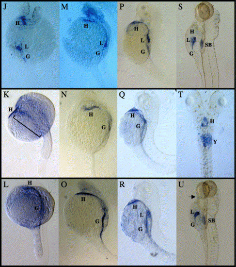
Whole-mount in situ hybridization analysis of GATA-4/5/6. (A–C) Dorsal views of five somite (12 hpf) embryos, anterior at the top, showing GATA-4 (A), GATA-5 (B) and GATA-6 (C) expression in the lateral plate mesoderm (LPM). (D–F) Dorsal views of 10 somite (14 hpf) embryos, anterior at the top, showing GATA-4 (D), GATA-5 (E) and GATA-6 (F) expression. All three GATA factors are expressed in the LPM. However, GATA-4 is also expressed in the tail bud (*) as previously reported in zebrafish (Griffin et al., 2000), GATA-5 expression is lower in the central portion of the LPM (denoted by a line) and GATA-6 is expressed posteriorly in the region medial to the stripes of the LPM (arrow) as previously shown in mouse (Morrisey et al., 1996). D is rotated slightly relative to E and F to show tail bud expression. (G–I) Dorsal views of embryos at 18 hpf, anterior at the top, showing GATA-4 (G), GATA-5 (H) and GATA-6 (I) expression in the cardiac progenitors migrating to the ventral aspect of the embryo. GATA-5 retains expression in posterior regions of the LPM (bracket) as shown previously in Xenopus (Jiang and Evans, 1996). (J–L) Dorsal views at 24 hpf, anterior at the top. GATA-4 (J), GATA-5 (K) and GATA-6 (L) expression can be seen in the fused heart tube. GATA-4 is also expressed in the liver, GATA-5 continues to have low levels of expression in the posterior LPM (bracket) and GATA-6 is expressed in a broad region of the gut tube. (M–O) Lateral views at 28 hpf, anterior at the top, showing GATA-4 (M), GATA-5 (N) and GATA-6 (O) expression at the beating heart stage. GATA-4 expression remains in the liver but now extends to more extensive regions of the gut. GATA-5 expression in the gut is just visible and restricted to the central portion. GATA-6 displays the most extensive pattern of gut expression but does not appear to be expressed at high levels in the liver at this stage. (P–R) Lateral views at 48 hpf, anterior at the top, showing GATA-4 (P), GATA-5 (Q) and GATA-6 (R) expression in the heart and gut tube. (S, U) Lateral and (T) ventral views at 6 days post fertilization, anterior at the top, showing GATA-4 (S) expression in the heart and gut, GATA-5 (T) expression in the heart, and GATA-6 (U) expression in the gut, heart and two of the posterior pharyngeal pouches (arrow). Stainings in the swim bladder in (S) and (U) and in the yolk in (T) are nonspecific staining artifacts. G = gut, H = heart, L = liver, SB = swim bladder and Y = yolk. |
| Genes: | |
|---|---|
| Fish: | |
| Anatomical Terms: | |
| Stage Range: | 5-9 somites to Day 6 |
Reprinted from Developmental Biology, 267(2), Heicklen-Klein, A., and Evans, T., T-box binding sites are required for activity of a cardiac GATA-4 enhancer, 490-504, Copyright (2004) with permission from Elsevier. Full text @ Dev. Biol.